. 2014 Jan 15;189(2):183-93.
doi: 10.1164/rccm.201306-1039OC.

Use of induced pluripotent stem cells to recapitulate pulmonary alveolar proteinosis pathogenesis

Affiliations

Use of induced pluripotent stem cells to recapitulate pulmonary alveolar proteinosis pathogenesis

Takuji Suzuki et al. Am J Respir Crit Care Med. .

Abstract

Rationale: In patients with pulmonary alveolar proteinosis (PAP) syndrome, disruption of granulocyte/macrophage colony-stimulating factor (GM-CSF) signaling is associated with pathogenic surfactant accumulation from impaired clearance in alveolar macrophages.

Objectives: The aim of this study was to overcome these barriers by using monocyte-derived induced pluripotent stem (iPS) cells to recapitulate disease-specific and normal macrophages.

Methods: We created iPS cells from two children with hereditary PAP (hPAP) caused by recessive CSF2RA(R217X) mutations and three normal people, differentiated them into macrophages (hPAP-iPS-Mφs and NL-iPS-Mφs, respectively), and evaluated macrophage functions with and without gene-correction to restore GM-CSF signaling in hPAP-iPS-Mφs.

Measurements and main results: Both hPAP and normal iPS cells had human embryonic stem cell-like morphology, expressed pluripotency markers, formed teratomas in vivo, had a normal karyotype, retained and expressed mutant or normal CSF2RA genes, respectively, and could be differentiated into macrophages with the typical morphology and phenotypic markers. Compared with normal, hPAP-iPS-Mφs had impaired GM-CSF receptor signaling and reduced GM-CSF-dependent gene expression, GM-CSF- but not M-CSF-dependent cell proliferation, surfactant clearance, and proinflammatory cytokine secretion. Restoration of GM-CSF receptor signaling corrected the surfactant clearance abnormality in hPAP-iPS-Mφs.

Conclusions: We used patient-specific iPS cells to accurately reproduce the molecular and cellular defects of alveolar macrophages that drive the pathogenesis of PAP in more than 90% of patients. These results demonstrate the critical role of GM-CSF signaling in surfactant homeostasis and PAP pathogenesis in humans and have therapeutic implications for hPAP.


Figures

Figure 1.
Figure 1.
Characterization of induced pluripotent stem cells (iPS cells) from hereditary pulmonary alveolar proteinosis (hPAP) and healthy individuals (NL). (A) Generation of patient/lung disease–specific iPS cells. Peripheral blood mononuclear cells (PBMCs) from healthy individuals or patients with hPAP were transduced to express iPS cell-inducing factors (indicated) and sequentially grown in specialized culture media and then on feeder cells to create cell clones as described in the Methods. Results for one iPS cell clone from a healthy individual (NL-iPS cells) and/or from a patient with hPAP homozygous for CSF2RAR217X mutations (hPAP-iPS cells) are shown. (B) iPS cell colony morphology. hPAP PBMCs, hPAP-iPS cell colonies, and human embryonic stem cell colonies (H9 hES) were examined by phase-contrast microscopy at low power (×10, ×5, ×5, left, center, right, respectively). Bars represent 200, 500, and 500 μm (left, center, and right, respectively). (C) Expression markers of pluripotency. Undifferentiated hPAP-iPS cell colonies were examined by immunofluorescence microscopy to detect protein markers of pluripotency (indicated) or cell nuclei (DAPI) (×20). Immunofluorescence analysis of NL-iPS cell colonies (not shown) was similar to that of hPAP-iPS cell colonies. (D) Teratoma formation assay analysis. Undifferentiated hPAP-iPS cell colonies were injected into immunodeficient mice permitting the formation of iPS cell–derived tumors, which were then removed and examined histopathologically. The pluripotency of the hPAP-iPS cells was confirmed by the formation of teratoma containing tissue elements derived from all three germ layers including respiratory epithelium (endoderm), cartilage (mesoderm), and neural epithelium (ectoderm) (hematoxylin and eosin). (E) Alkaline phosphatase activity. Undifferentiated hPAP-iPS cell colonies were evaluated for alkaline phosphatase, a pluripotent cell marker, by routine histochemical methods as described in the Methods. One 35-mm-diameter well containing multiple positively stained hPAP-iPS cell colonies is shown. Histochemical analysis of NL-iPS cell colonies (not shown) was similar to that of hPAP-iPS cell colonies. (F) Karyotype analysis. Undifferentiated hPAP-iPS cell colonies were evaluated by routine clinical karyotyping analysis, which confirmed a normal complement of morphologically normal-appearing chromosomes. (G) CSF2RA gene nucleotide sequencing. Routine polymerase chain reaction–based nucleotide sequencing of genomic DNA confirmed homozygous normal or R217X sequences for the CSF2RA gene in NL-iPS cell and hPAP-iPS cell colonies, respectively.
Figure 2.
Figure 2.
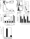
Directed differentiation of macrophages from hereditary pulmonary alveolar proteinosis (hPAP) induced pluripotent stem cells (iPS cells) and iPS cell clones from healthy individuals (NL). Undifferentiated hPAP-iPS or NL-iPS cell colonies were differentiated into macrophages (hPAP-iPS-Mφs or NL-iPS-Mφs, respectively) as described in the Methods and the online supplement. (A) Generation of iPS cell–derived macrophages. Independent iPS cell clones were cultured on OP9 mouse bone marrow stromal feeder cells to direct multipotent lineage commitment and expanded in suspension culture in media containing macrophage colony–stimulating factor (M-CSF) and granulocyte-macrophage colony–stimulating factor (GM-CSF) to expand myeloid lineage cells. Immunomagnetic separation was used to isolate CD45+/CD235a/CD41a macrophage precursors, which were expanded and differentiated in media containing M-CSF and GM-CSF. (B) Evaluation of the myeloid cell expansion efficiency. The efficiency of myeloid lineage cell expansion from iPS cells was evaluated for iPS cell clones from three different healthy individuals (NL-1, -2, -3) and two patients with hPAP (hPAP-1, -2) by measuring the percentage of CD45+/CD235a/CD41a cells. Each symbol indicates a separate determination. (C) Morphology of live hPAP-iPS cell-derived macrophages. Cells were examined by phase-contrast microscopy (original photomicrograph ×20). (D) Morphology of sedimented hPAP-iPS cell–derived macrophages. Cells were sedimented onto slides (Cytospin), stained (Diff-Quick), and examined by light microscopy (original photomicrograph ×20). (E). Phenotypic marker analysis. Cells were immunostained with specific antibodies to various phenotypic markers (shaded histograms) or isotype control antibodies (clear histograms) as indicated and evaluated by flow cytometry as described in the Methods. Shown is gating strategy (red ovals in top panels) used and phenotypic assessment (lower panels) for one normal (NL-1) and one hPAP (hPAP-1) clone. The percentage (±SEM) of differentiated macrophages that were CD68+ was 98 ± 0.8% for all clones evaluated (NL-1, -2, -3, and hPAP-1, -2). Additional results for NL-2, NL-3, and hPAP-2 are included in Figure E3.
Figure 3.
Figure 3.
Granulocyte-macrophage colony–stimulating factor (GM-CSF) receptor function in hereditary pulmonary alveolar proteinosis (hPAP) induced pluripotent stem cells (iPS cells) and healthy individuals (NL)–iPS cell–derived macrophages. (A) Cell/receptor-mediated GM-CSF clearance. GM-CSF was added at time zero to the media of culture dishes containing NL-iPS-Mφs (closed circles), hPAP-iPS-Mφs (open circles) or media without cells (gray diamonds). At subsequent times, GM-CSF in the culture media was measured by ELISA and the amount remaining was compared with the initial concentration. Using analysis of variance, subsequent values differing significantly from initial values are indicated (*P < 0.001). (B) Receptor-mediated signal transducer and activator of transcription 5 (STAT5) phosphorylation. NL-iPS-Mφs or hPAP-iPS-Mφs were incubated for 30 minutes with or without 10 ng/ml GM-CSF as indicated and then phosphorylated STAT5 immunostaining and flow cytometry were used to measure the STAT5 phosphorylation index (STAT5-PI) as described in the Methods. Each symbol indicates a separate expansion of iPS cell–derived Mφs from one clone from one of the three normal individuals and two patients with hPAP (different symbol types represent results for cells from each individual; NL = upright triangles, downward triangles, squares; hPAP = circles, diamonds). (C) CSF-stimulated cellular proliferation. NL-iPS-Mφs or hPAP-iPS-Mφs were cultured with or without GM-CSF or M-CSF at the indicated concentrations for 72 hours and then cellular proliferation was measured by the XTT assay as described in the Methods. Bars represent the mean (±SEM) of four determinations on one clone each from NL-1 and hPAP-1. The cellular proliferation rate at various CSF concentrations was compared with that of unexposed cells by analysis of variance; values differing significantly are indicated (*P < 0.001). (D) Gene expression in hPAP-iPS-Mφs and NL-iPS-Mφs. Levels of mRNA transcript for several key genes regulated by GM-CSF in macrophages were measured in total RNA from NL-iPS-Mφs (NL) or hPAP-iPS-Mφs (hPAP) after conversion to cDNA by quantitative real-time polymerase chain reaction as described in the Methods. Transcript levels were normalized to the mean value in NL-iPS-Mφs for each gene. Bars represent the mean (±SEM) of four determinations on one clone each from NL-1 and hPAP-1. Comparisons between hPAP-iPS-Mφs and NL-iPS-Mφs were made using Student t test; values differing significantly are indicated (*P < 0.05). (E) Host defense-related proinflammatory signaling by hPAP-iPS-Mφs and NL-iPS-Mφs. NL-iPS-Mφs or hPAP-iPS-Mφs were cultured with or without LPS (100 ng/ml) for 24 hours and then tumor necrosis factor (TNF)-α released into the medium was measured by ELISA. Bars represent the mean (±SEM) of four determinations on one clone each from NL-1 and hPAP-1. TNF-α levels in unstimulated cells (*) were minimal (90.90 ± 8.56 and 58.58 ± 22.14 pg/ml in NL- and hPAP-iPS-Mφs, respectively). LPS stimulated TNF-α release was markedly reduced in hPAP-iPS-Mφs compared with NL-iPS-Mφs (Mann-Whitney rank sum test, P < 0.05).
Figure 4.
Figure 4.
Impaired pulmonary surfactant clearance in hereditary pulmonary alveolar proteinosis (hPAP) induced pluripotent stem cells (iPS cells) Mφs and correction by gene transfer-mediated restoration of granulocyte-macrophage colony–stimulating factor (GM-CSF) receptor function. (A) Schematic of evaluation of cells before and after correction of GM-CSF signaling. Lentiviral-mediated transfer of the normal CSF2RA cDNA expressing the GM-CSF receptor α into hPAP-iPS cells was performed as described in the Methods and the gene-corrected cells (or uncorrected hPAP-iPS cells or NL-iPS cells) were differentiated into macrophages as shown in Figure 2 and evaluated as indicated. (B) Oil red O staining of iPS cell–derived macrophages. NL-iPS-Mφs, hPAP-iPS-Mφs, or gene-corrected hPAP-iPS-Mφs (hPAP-iPS-Mφs+LV-hCSF2RA) (top panels) were exposed to surfactant for 24 hours (middle panels) and then washed with phosphate-buffered saline and cultured 24 hours more in the absence of surfactant (lower panels). Cells were sedimented onto slides (Cytospin), stained (oil red O) and examined by light microscopy and photographed. (C) Evaluation of CD116 on gene-corrected hPAP-iPS-Mφs. Cells were immunostained with anti-human CD116 antibodies (shaded) or isotype control antibodies (open) and cell surface CD116 levels were evaluated by measuring fluorescence by flow cytometry. LV-GFP and LV-hCSF2RA indicate control and hCSF2RA vectors, respectively. (D) GM-CSF receptor-signaling in gene-corrected hPAP-iPS-Mφs. Cells were incubated for 30 minutes with or without 10 ng/ml GM-CSF, immunostained with anti–phospho–signal transducer and activator of transcription 5 (STAT5) specific antibody, and then intracellular phospho-STAT5 levels were measured by flow cytometry as described in the Methods. (E) Measurement of lipid accumulation in iPS cell–derived macrophages. Macrophages were cultured in the presence and then absence of surfactant followed by washing and staining with oil red O as described (Figure 4B) and evaluated by light microscopy using a visual grading scale (the oil red O staining index) to measure the degree of staining as previously described (8). Bars represent the mean (±SEM) oil red O staining score for 10 high-power fields for one clone each from NL-1 and hPAP-1. Results shown are from one of three representative experiments with these clones. Similar results were obtained in three experiments with additional clones (see Figure E4).

Comment in

Similar articles

Cited by

References

    1. Suzuki T, Sakagami T, Rubin BK, Nogee LM, Wood RE, Zimmerman SL, Smolarek T, Dishop MK, Wert SE, Whitsett JA, et al. Familial pulmonary alveolar proteinosis caused by mutations in CSF2RA. J Exp Med. 2008;205:2703–2710. - PMC - PubMed
    1. Suzuki T, Sakagami T, Young LR, Carey BC, Wood RE, Luisetti M, Wert SE, Rubin BK, Kevill K, Chalk C, et al. Hereditary pulmonary alveolar proteinosis: pathogenesis, presentation, diagnosis, and therapy. Am J Respir Crit Care Med. 2010;182:1292–1304. - PMC - PubMed
    1. Martinez-Moczygemba M, Doan ML, Elidemir O, Fan LL, Cheung SW, Lei JT, Moore JP, Tavana G, Lewis LR, Zhu Y, et al. Pulmonary alveolar proteinosis caused by deletion of the GM-CSFRalpha gene in the X chromosome pseudoautosomal region 1. J Exp Med. 2008;205:2711–2716. - PMC - PubMed
    1. Suzuki T, Maranda B, Sakagami T, Catellier P, Couture CY, Carey BC, Chalk C, Trapnell BC. Hereditary pulmonary alveolar proteinosis caused by recessive CSF2RB mutations. Eur Respir J. 2011;37:201–204. - PubMed
    1. Tanaka T, Motoi N, Tsuchihashi Y, Tazawa R, Kaneko C, Nei T, Yamamoto T, Hayashi T, Tagawa T, Nagayasu T, et al. Adult-onset hereditary pulmonary alveolar proteinosis caused by a single-base deletion in CSF2RB. J Med Genet. 2011;48:205–209. - PubMed

MeSH terms

Substances

Supplementary concepts