. 2013 Jan 23;1(1):5.
doi: 10.1186/2049-3002-1-5.

Activated lymphocytes as a metabolic model for carcinogenesis

Affiliations

Activated lymphocytes as a metabolic model for carcinogenesis

Andrew N Macintyre et al. Cancer Metab. .

Abstract

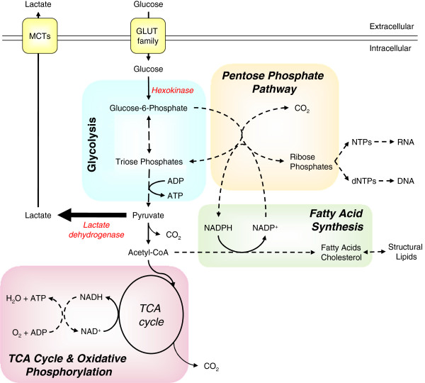
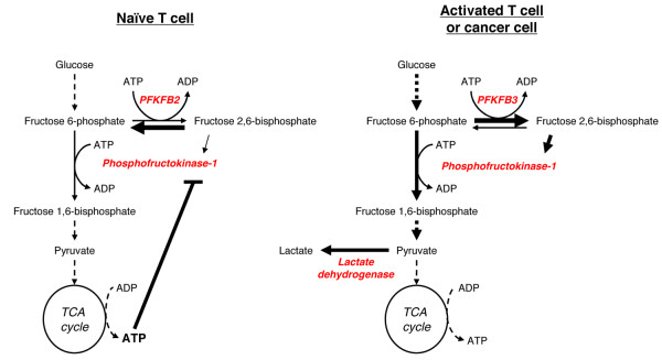
Metabolic reprogramming is a key event in tumorigenesis to support cell growth, and cancer cells frequently become both highly glycolytic and glutamine dependent. Similarly, T lymphocytes (T cells) modify their metabolism after activation by foreign antigens to shift from an energetically efficient oxidative metabolism to a highly glycolytic and glutamine-dependent metabolic program. This metabolic transition enables T cell growth, proliferation, and differentiation. In both activated T cells and cancer cells metabolic reprogramming is achieved by similar mechanisms and offers similar survival and cell growth advantages. Activated T cells thus present a useful model with which to study the development of tumor metabolism. Here, we review the metabolic similarities and distinctions between activated T cells and cancer cells, and discuss both the common signaling pathways and master metabolic regulators that lead to metabolic rewiring. Ultimately, understanding how and why T cells adopt a cancer cell-like metabolic profile may identify new therapeutic strategies to selectively target tumor metabolism or inflammatory immune responses.


Figures

Figure 1
Figure 1
Major metabolic fates of glucose in highly proliferative cells. Glucose is taken into the cell by GLUT family transporters and then phosphorylated by hexokinases, trapping it within the cell as glucose-6-phosphate (G6P). G6P can be catabolized via glycolysis or used as a carbon donor for the synthesis of riboses via the pentose phosphate pathway (PPP). Catabolized G6P generates pyruvate plus small quantities of ATP, with much of the resultant pyruvate being converted to lactate by lactate dehydrogenase and then secreted through mono-carboxylic transporters (MCT). The remaining pyruvate is converted to acetyl-CoenzymeA (acetyl-CoA) by pyruvate dehydrogenase and used either as fuel for ATP production via the tri-carboxylic acid (TCA) cycle and oxidative phosphorylation or converted to fatty acids to generate structural lipids. At various points during glycolysis and the TCA cycle reaction intermediates can be removed to provide carbon for amino acid biosynthesis (not shown).
Figure 2
Figure 2
T cell activation results in metabolic reprogramming. Naïve T cells have an oxidative metabolism, using glucose, glutamine, and fatty acids as fuel sources. The majority of ATP is generated via oxidative phosphorylation. Following activation by stimulation of the T cell receptor and co-receptors, the cells adopt a metabolic profile that resembles the metabolism of many cancer cells, consuming large quantities of both glucose and glutamine but performing relatively little oxidative phosphorylation. The majority of glucose-derived carbon is secreted as lactate, with the remainder being used for biosynthesis.
Figure 3
Figure 3
Glycolytic isozyme switching promotes high rates of glycolysis. Activated T cells, cancer cells and other highly proliferative cells express different glycolytic isozymes in comparison to quiescent cells, increasing glycolytic flux. One key step in glycolysis is the phosphorylation of fructose 6-phosphate by phosphofructokinase-1 (PFK-1). PFK-1 is allosterically activated by fructose 2,6-bisphosphate and allosterically inhibited by ATP. Both activated T cells and tumor cells express isoform 3 of the bifunctional enzyme 6-phosphofructo-2-kinase/fructose-2,6-bisphosphatase (PFKFB). In contrast, naïve T cells express PFKFB isoform 2. PFKFB3 differs from PFKFB2 in that it has low phosphatase activity, leading to the accumulation of fructose 2,6-bisphosphate and localized depletion of ATP. This results in increased PFK-1 activity and higher rates of glycolysis.

References

    1. Khorana HG. Chemical Biology; Selected Papers of H. Gobind Khorana. Singapore: World Scientific Publishing Co; 2000.
    1. Gerriets VA, Rathmell JC. Metabolic pathways in T cell fate and function. Trends Immunol. 2012;33:168–173. doi: 10.1016/j.it.2012.01.010. - DOI - PMC - PubMed
    1. Anderson G, Lane PJ, Jenkinson EJ. Generating intrathymic microenvironments to establish T-cell tolerance. Nat Rev Immunol. 2007;7:954–963. doi: 10.1038/nri2187. - DOI - PubMed
    1. Lunt SY, Vander Heiden MG. Aerobic glycolysis: meeting the metabolic requirements of cell proliferation. Annu Rev Cell Dev Biol. 2011;27:441–464. doi: 10.1146/annurev-cellbio-092910-154237. - DOI - PubMed
    1. Deberardinis RJ, Lum JJ, Thompson CB. Phosphatidylinositol 3-kinase-dependent modulation of carnitine palmitoyltransferase 1A expression regulates lipid metabolism during hematopoietic cell growth. J Biol Chem. 2006;281:37372–37380. doi: 10.1074/jbc.M608372200. - DOI - PubMed