Review
. 2014 Jun;20(3):272-90.
doi: 10.1177/1073858413510044. Epub 2013 Nov 25.

Spontaneous Network Activity and Synaptic Development

Affiliations
Review

Spontaneous Network Activity and Synaptic Development

Daniel Kerschensteiner. Neuroscientist. 2014 Jun.

Abstract

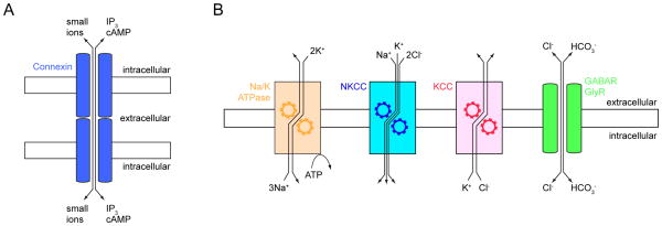
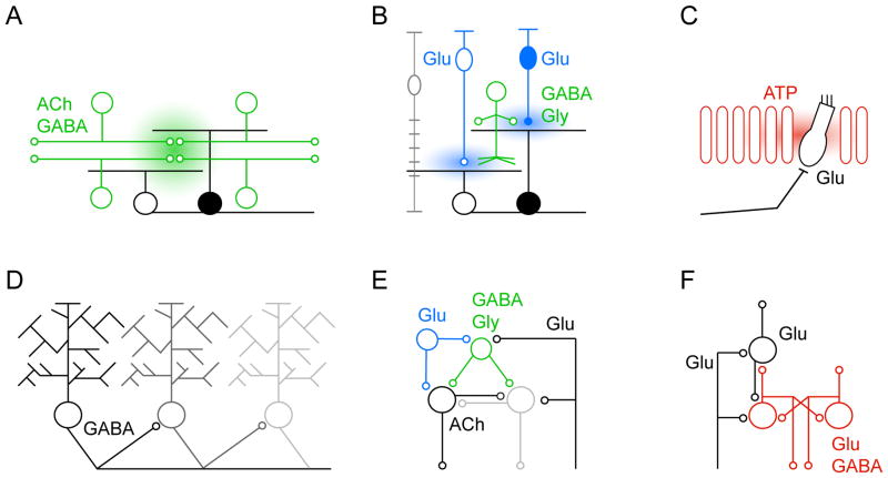
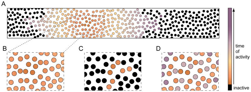
Throughout development, the nervous system produces patterned spontaneous activity. Research over the past two decades has revealed a core group of mechanisms that mediate spontaneous activity in diverse circuits. Many circuits engage several of these mechanisms sequentially to accommodate developmental changes in connectivity. In addition to shared mechanisms, activity propagates through developing circuits and neuronal pathways (i.e., linked circuits in different brain areas) in stereotypic patterns. Increasing evidence suggests that spontaneous network activity shapes synaptic development in vivo Variations in activity-dependent plasticity may explain how similar mechanisms and patterns of activity can be employed to establish diverse circuits. Here, I will review common mechanisms and patterns of spontaneous activity in emerging neural networks and discuss recent insights into their contribution to synaptic development.

Keywords: circuit mechanisms; connectivity; patterned activity; plasticity; synaptogenesis; waves.


Conflict of interest statement

Declaration of Conflicting Interests: The author declares no potential conflicts of interest with respect to the research, authorship, and/or publication of this article.

Figures

Figure 1
Figure 1
Gap junctions and chloride homeostasis. (A) Aligned connexin hemichannels (blue) in adjacent plasma membranes provide a direct pathway for small ions and signaling molecules between neurons. (B) Na+ and K+ concentration gradients established by the Na+/K+ ATPase (Na/K ATPase, orange) provide the energy for the two transport systems that control neuronal [Cl]i. Na+-K+-2Cl- cotransporters (NKCCs, blue) import Cl, whereas K+-Cl cotransporters (KCCs, red) export Cl. Cl flows through GABA and glycine receptors (GABAR and GlyR, green) down its electrochemical gradient. Cl efflux depolarizes and Cl influx hyperpolarizes a neuron.
Figure 2
Figure 2
Diverse circuits generate patterned spontaneous activity. (A) Reciprocally connected starburst amacrine cells (SACs, green) initiate and propagate stage II waves in the retina, activating neighboring ON (white) and OFF (black) retinal ganglion cells (RGCs) simultaneously via extrasynaptic diffusion of ACh (and GABA). (B) Glutamate spillover and gap junctions mediate communication between neighboring ON bipolar cells (ON BCs) (open, blue) and laterally spread activity during stage retinal III waves. In addition, ON BCs activate ON RGCs (white) and diffuse ACs (green), which crossover inhibit OFF BCs (closed, blue) and delay their release of glutamate. Sequential glutamate release from ON and OFF BCs desynchronizes the activity of neighboring ON and OFF (black) RGCs during stage III waves. Glutamate uptake by Müller glia (gray) limits vertical spillover and maintains separation between ON and OFF circuits. (C) Support cells (red) in Kölliker’s organ of the developing cochlea generate waves of extracellular ATP, which synchronize the spontaneous spike bursts of nearby inner hair cells (IHCs, white). IHCs in turn release glutamate and activate spiral ganglion neurons. (D) In the developing cerebellum, spontaneous waves of activity travel along chains of PCs. Activity is transmitted in a directional manner established by asymmetric axon collateral. (E) Abundantly connected networks of GABA- and glycinergic interneurons (green, Renshaw cells), glutamatergic interneurons (blue), cholinergic motorneurons and glutamatergic proprioceptive inputs generate spontaneous activity patterns in the developing spinal cord. (F) Subplate neurons (SPNs, red) are a hub of developing thalamocortical circuits. SPNs receive input from thalamus, are reciprocally connected and receive feedback from the developing cortical plate. In addition to feedforward connections particularly with layer 4 neurons (white), SPNs provide feedback to thalamus. The resulting loops can generate activity and amplify activity originating in the sensory periphery (e.g. retina).
Figure 3
Figure 3
Characteristic patterns of neuronal activation during different spontaneous network events. (A) The large scale propagation of activity during spreading waves is illustrated by color-coding the time of cellular recruitment. (B) An excerpt from a smaller region shows homogenous and synchronous activation of nearby neurons. (C) By contrast, restricted clusters of neurons are activated during synchronized assemblies. (D) In some instances (e.g. stage III retinal waves), different types of neighboring neurons are recruited sequentially into spreading waves (i.e. precise asynchrony).

References

    1. Abreu-Villaca Y, Filgueiras CC, Manhaes AC. Developmental aspects of the cholinergic system. Behav Brain Res. 2011;221(2):367–78. - PubMed
    1. Ackman JB, Burbridge TJ, Crair MC. Retinal waves coordinate patterned activity throughout the developing visual system. Nature. 2012;490(7419):219–25. - PMC - PubMed
    1. Adelsberger H, Garaschuk O, Konnerth A. Cortical calcium waves in resting newborn mice. Nat Neurosci. 2005;8(8):988–90. - PubMed
    1. Ahrens MB, Orger MB, Robson DN, Li JM, Keller PJ. Whole-brain functional imaging at cellular resolution using light-sheet microscopy. Nat Methods. 2013;10(5):413–20. - PubMed
    1. Akemann W, Knopfel T. Interaction of Kv3 potassium channels and resurgent sodium current influences the rate of spontaneous firing of Purkinje neurons. J Neurosci. 2006;26(17):4602–12. - PMC - PubMed

LinkOut - more resources