. 2013 Dec;4(12):2366-82.
doi: 10.18632/oncotarget.1424.

Ovarian cancer stem-like cells show induced translineage-differentiation capacity and are suppressed by alkaline phosphatase inhibitor

Affiliations

Ovarian cancer stem-like cells show induced translineage-differentiation capacity and are suppressed by alkaline phosphatase inhibitor

Kuei-Chun Liu et al. Oncotarget. 2013 Dec.

Abstract

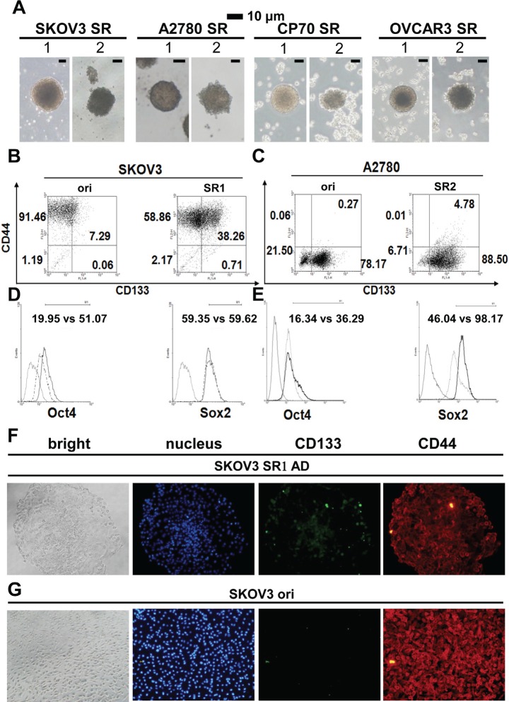
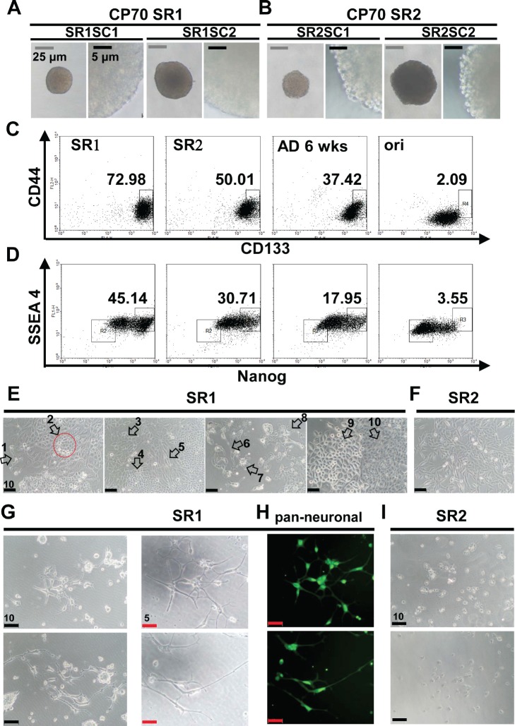
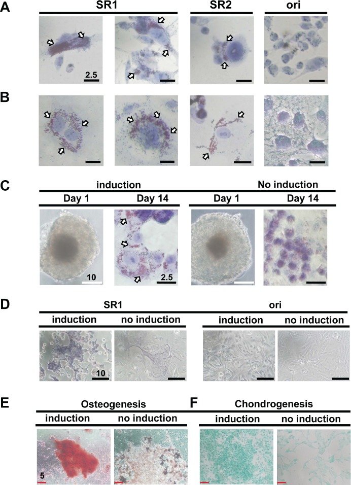
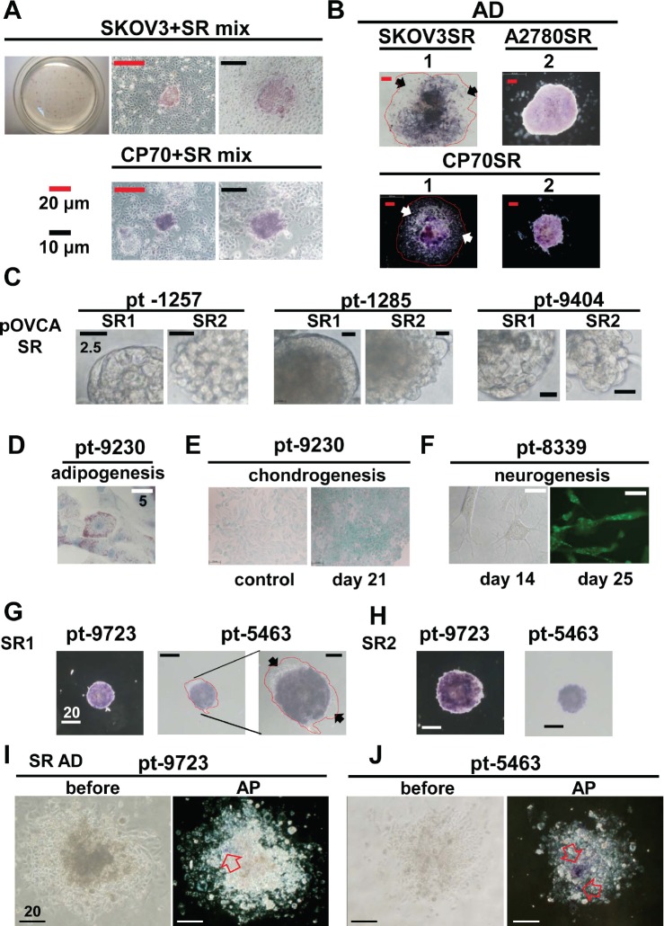
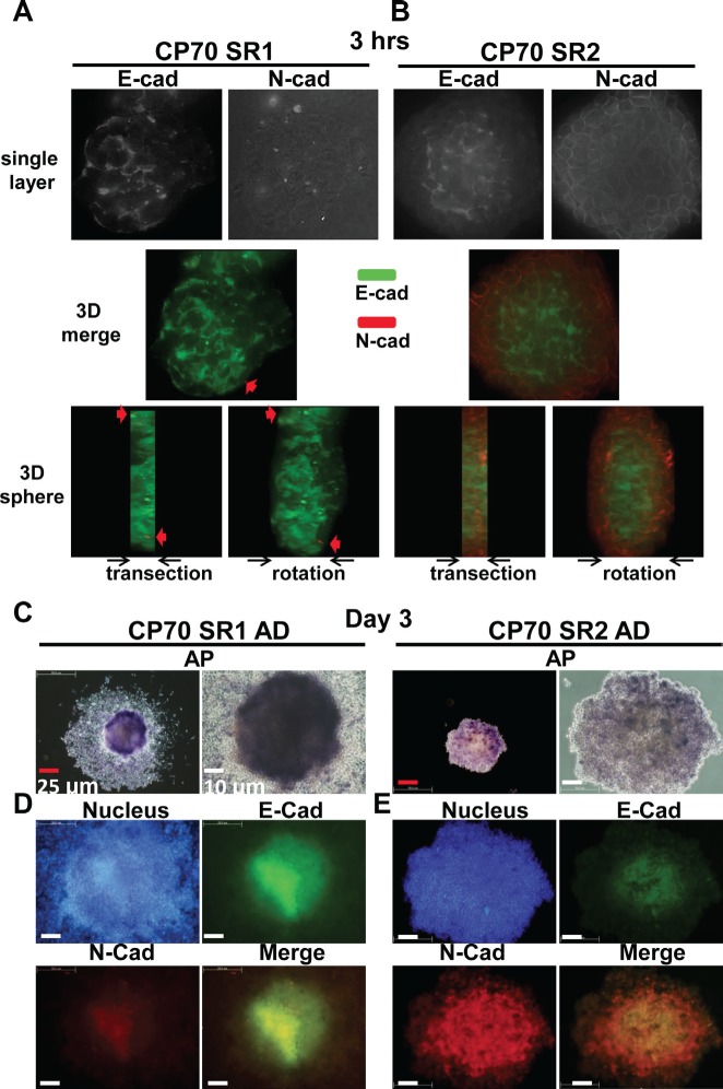
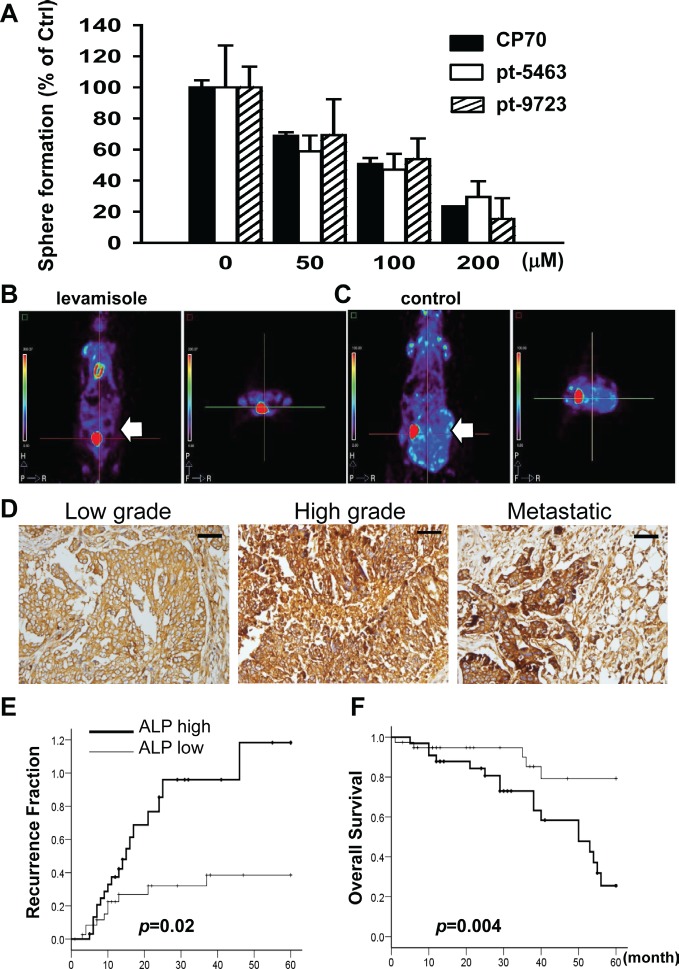
Spheroid formation is one property of stem cells-such as embryo-derived or neural stem cells-that has been used for the enrichment of cancer stem-like cells (CSLCs). However, it is unclear whether CSLC-derived spheroids are heterogeneous or whether they share common embryonic stemness properties. Understanding these features might lead to novel therapeutic approaches. Ovarian carcinoma is a deadly disease of women. We identified two types of spheroids (SR1 and SR2) from ovarian cancer cell lines and patients' specimens according to their morphology. Both types expressed stemness markers and could self-renew and initiate tumors when a low number of cells were used. Only SR1 could differentiate into multiple-lineage cell types under specific induction conditions. SR1 spheroids could differentiate to SR2 spheroids through epithelial-mesenchymal transition. Alkaline phosphatase (ALP) was highly expressed in SR1 spheroids, decreased in SR2 spheroids, and was absent in differentiated progenies in accordance with the loss of stemness properties. We verified that ALP can be a marker for ovarian CSLCs, and patients with greater ALP expression is related to advanced clinical stages and have a higher risk of recurrence and lower survival rate. The ALP inhibitor, levamisole, disrupted the self-renewal of ovarian CSLCs in vitro and tumor growth in vivo. In summary, this research provides a plastic ovarian cancer stem cell model and a new understanding of the cross-link between stem cells and cancers.This results show that ovarian CSLCs can be suppressed by levamisole. Our findings demonstrated that some ovarian CSLCs may restore ALP activity, and this suggests that inhibition of ALP activity may present a new opportunity for treatment of ovarian cancer.


Conflict of interest statement

The authors declare no potential conflicts of interest.

Figures

Figure 1
Figure 1. Morphology and stemness properties of ovarian cancer cell spheroids
(A) Two different types of spheroids (SR1 and SR2) were enriched in various ovarian cancer cell lines. (B, C) The CD44+CD133+ signal increased in both SR1 and SR2 cells derived from SKOV3 and A2780 cells, whereas CD44+CD133 and CD44CD133+ were expressed in the original SKOV3 and A2780 cells, respectively. The expression of CD133 and OCT4 was higher in SKOV3SR1 cells (B, D), whereas the expression of OCT4 and SOX2 was higher in A2780SR2 cells (C, E). (F, G) Expression of CD133 and CD44 was compared between adhesive SKOV3-derived SR1 and the original cells. (F) After adhesive culture for 24 hours, the expression of CD133 decreased in adhesive SKOV3-derived SR1 from the center site to the periphery, but CD133 in scattered SR1 cells was still detectable in the periphery. (G) Almost all SKOV3 original cells expressed CD44 but not CD133.
Figure 2
Figure 2. Characterization of single-cell clones isolated from CP70 SR1 and SR2 spheroids
(A, B) Single cell-derived SR1 and SR2 clones are referred to as SR1SC and SR2SC, respectively. The surface of SR1SC (A) was smooth regardless of the size, whereas SR2SC (B) was morula like. (C, D) Stemness-associated markers were detected in single cell-derived SR1, SR2, and SR cells 6 weeks after differentiation, and in the original cells. (C) The population of CD133+/CD44+ cells was the highest in SR1 followed by SR2, differentiated SR cells, and the original cells. (D) Expression of both the stemness-associated marker NANOG and hESC-related marker SSEA4 was increased in the order SR1, SR2, and SR differentiated cells. SSEA4 expression was lower in CP70 original cells than in their counterpart SR cells. (E) Dissociated SR1SC cells were induced to differentiate for 14 days. At least 10 types of morphology (white arrows) with a neuron-like phenotype were observed in different fields of view, whereas CP70SR2 cells differentiated only to a simple and tumor-like morphology (F). Neuron-like morphology was observed only in CP70SR1 cells (G), and expression of multiple neuronal markers was detected by immunostaining using anti-pan-neuronal antibodies (H). However, most CP70SR2 cells were dead when treated under the condition described above for 16 days (I).
Figure 3
Figure 3. SR1 cells derived from various ovarian cancer cell lines show multiple differentiation potency
(A, B) SR1, SR2, and their original counterpart cells from CP70 (A) and SKOV3 (B) cells were induced by adipogenesis-inducing medium for 14 days and stained with oil red O solution. Only SR1 and SR2 cells, but not the original cells, displayed lipid droplets in the cell body (white arrows). (C) Two independent clones of SR1 cells derived from OVCAR3 cells were seeded directly onto collagen-coating culture dishes in either adipogenesis-inducing medium or SR culture medium. Oil droplets were observed only in SR1 cells cultured in adipogenesis-inducing medium. (D) The osteogenesis capacity of SKOV3-derived SR1 cells and its original counterpart cells were examined. Only SR1 cells cultured in osteogenesis-inducing medium displayed osteocyte morphology with the osteogenic marker, ALP activity, 14 days after induction. Differentiated CP70SR1 were induced by osteogenesis (E) and chondrogenesis (F) and detected by specific staining. Undifferentiated CP70SR1 without induction retained a tumor-like morphology and showed no signals.
Figure 4
Figure 4. Stemness-associated marker ALP increased in spheroid cells derived from ovarian cancer cell lines and patients' tissues
(A) ALP activity was assessed in spheroids and aggregated counterpart human SKOV3 and CP70 cells 18 h after attachment to the culture plate. ALP activity was detected only in spheroids in both cell lines. (B) ALP activity was upregulated in spheroids from three different cell lines, compared with their differentiated forms in cells that were allowed to attach to the culture plate in standard medium for 3–5 days. The red line encircles the extended region of adhesive SR1 cells, and arrows indicate ALP-negative differentiated SR1 cells. (C) SR1 and SR2 cell lines were isolated from ascites fluids from patients with ovarian cancers. Most spheroids displayed SR2 morphology. (D–F) SR cells isolated from patients' tissue were enriched and passaged three times for further assays. (D) After adipogenesis induction, SR cells could differentiate into adipocytes and displayed oil droplets in the cell bodies. (E) Alcian blue staining confirmed that isolated SR cells showed chondrogenic morphology and displayed the chondrocyte-specific marker 21 days after the induction of chondrogenesis. (F) Isolated SR cells exhibited a neuron-like morphology 14 days after induction and expressed αinternexin 21 days after induction. Patient specimens (solid tumor)-derived SR1 (G) and SR2 (H) cells were cultured in standard culture dishes for 6 h, and ALP activity was measured. (G) ALP activity was detectable only in undifferentiated SR cells and not in differentiated cells (black arrows). The red line encircles the extended region of adhesive SR1 cells. (I, J) When cultured in standard medium under adhesion conditions, after 7 days, ALP activity decreased significantly in differentiated SR cells derived from two individual patients. The left panel shows a bright field view of differentiated SR cells before ALP activity staining; the right panel shows the region of remaining ALP activity in the differentiated SR cells (open arrow).
Figure 5
Figure 5. Differences in ALP activity and expression of E-cadherin (E-cad) and N-cadherin (N-cad) between SR1 and SR2 cells
Entire spheroids of CP70SR1 (A) and CP70SR2 (B) were seeded into culture dishes for 3 hours and stained with combined antibodies against E-cad and N-cad (red arrow), and protein expression was analyzed by three-dimensional dissection using the DeltaVision system. E-cadherin was expressed throughout the entire SR1, only in the center of SR2, and N-cad was expressed only at the border. (C) After differentiation, SR1 cells grown under the adhesion condition showed a different pattern of ALP activity from the center to the periphery. SR2 cells showed weaker adhesion ability, and ALP activity was detectable only in certain regions. (D, E) The expression and location of E-cad and N-cad differed significantly between SR1 and SR2. The expression of E-cad and N-cad decreased from undifferentiated cells (center) to differentiated cells (boundary) in both SR1 and SR2. (D) E-cadherin was expressed mainly in the center of adhesive SR1, whereas the expression of N-cad was weaker at the same site. (E) In adhesive SR2, N-cad was expressed in most cells but with different strength, whereas E-cad was detectable only in the center.
Figure 6
Figure 6. Inhibition of ALP activity by levamisole interferes with the formation of spheroids and tumors in ovarian CSLCs
(A) Levamisole inhibited spheroid formation of dissociated single CP70SR1 cells and specimen-derived SR1 cells. The inhibitory efficacy of levamisole differed between spheroids isolated from different patients. For in vivo analysis of the effect of levamisole on ovarian CSLCs, mice were implanted peritoneally with CP70 cells and then given oral levamisole (B) or placebo (C). Tumor volume was significantly decreased in levamisole-treated mice as analyzed using a micro-PET image system. (D) ALP expression was associated with serous type and a high grade of EOC. In addition, metastatic cancer with the greatest expression of ALP indicated that ALP was related with advanced stage of EOC. Kaplan–Meier analysis of the probability of recurrence (E) and overall survival rate (F) in ovarian cancer patients stratified according to ALP expression (n = 73).

Similar articles

Cited by

References

    1. Pastrana E, Silva-Vargas V, Doetsch F. Eyes wide open: a critical review of sphere-formation as an assay for stem cells. Cell Stem Cell. 2011;8(5):486–498. - PMC - PubMed
    1. Frank NY, Schatton T, Frank MH. The therapeutic promise of the cancer stem cell concept. JClinInvest. 2010;120(1):41–50. - PMC - PubMed
    1. Valent P, Bonnet D, De Maria R, Lapidot T, Copland M, Melo JV, Chomienne C, Ishikawa F, Schuringa JJ, Stassi G, Huntly B, Herrmann H, Soulier J, Roesch A, Schuurhuis GJ, Wohrer S, et al. Cancer stem cell definitions and terminology: the devil is in the details. Nature reviews Cancer. 2012;12(11):767–775. - PubMed
    1. Vermeulen L, Sprick MR, Kemper K, Stassi G, Medema JP. Cancer stem cells--old concepts, new insights. Cell death and differentiation. 2008;15(6):947–958. - PubMed
    1. Bapat SA, Mali AM, Koppikar CB, Kurrey NK. Stem and progenitor-like cells contribute to the aggressive behavior of human epithelial ovarian cancer. Cancer Res. 2005;65(8):3025–3029. - PubMed

Publication types

MeSH terms

Substances