. 2013 Nov;25(11):4525-43.
doi: 10.1105/tpc.113.118463. Epub 2013 Nov 26.

Pollen tube growth regulation by free anions depends on the interaction between the anion channel SLAH3 and calcium-dependent protein kinases CPK2 and CPK20

Affiliations

Pollen tube growth regulation by free anions depends on the interaction between the anion channel SLAH3 and calcium-dependent protein kinases CPK2 and CPK20

Timo Gutermuth et al. Plant Cell. 2013 Nov.

Abstract

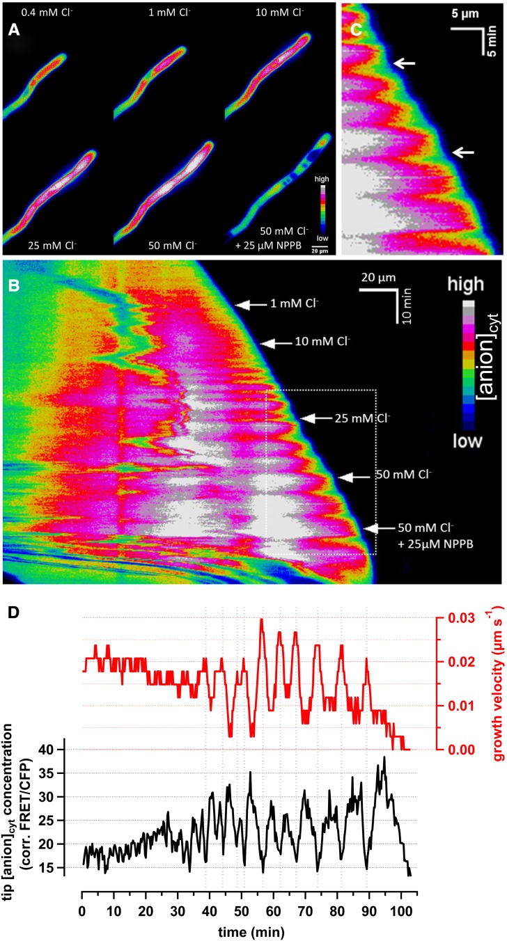
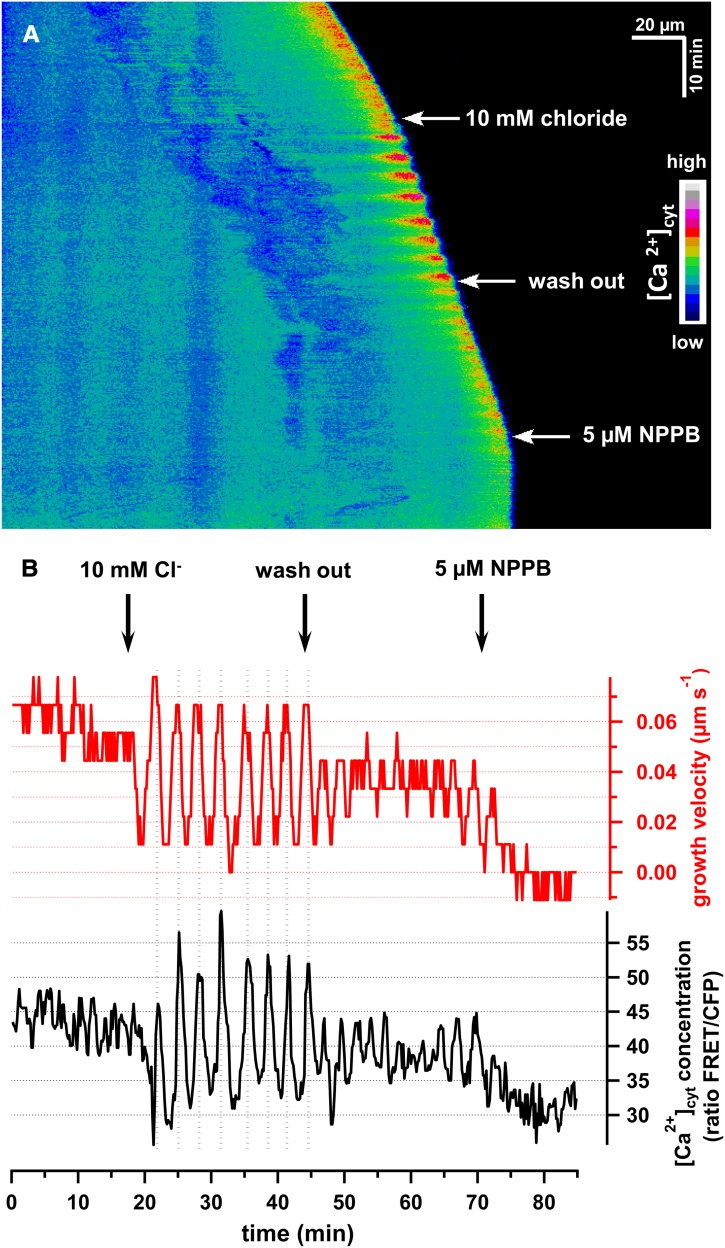
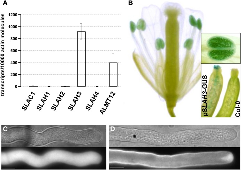
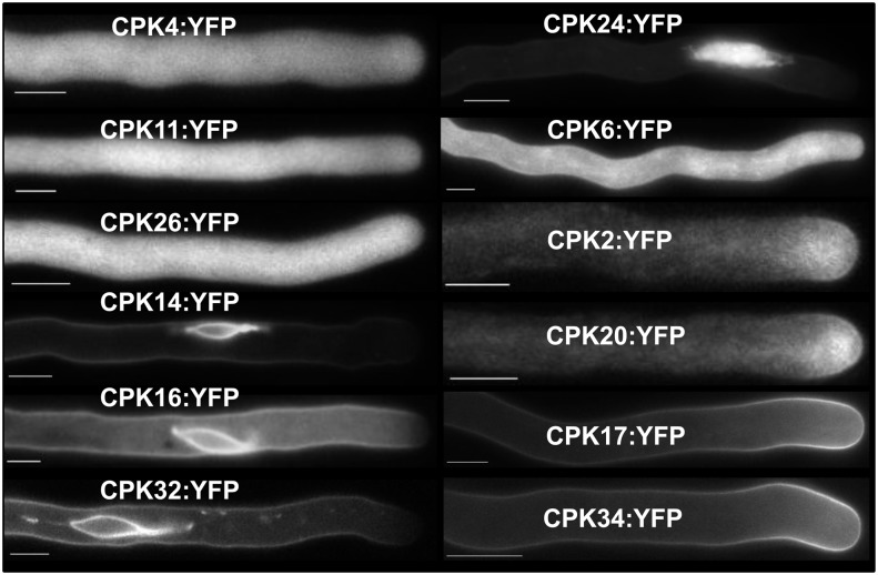
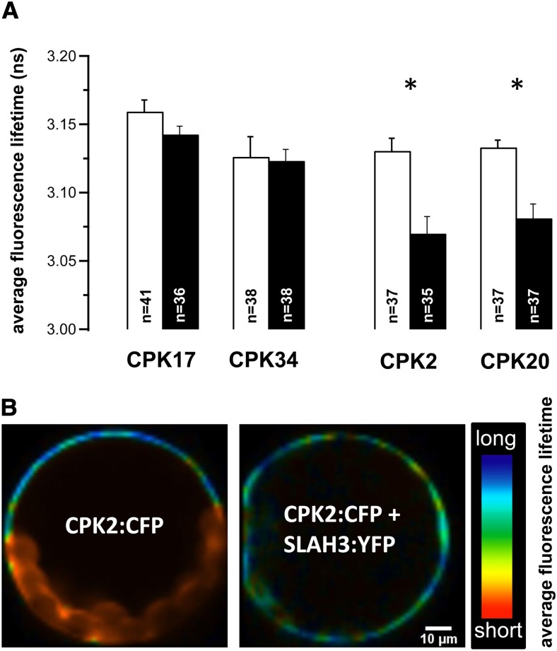
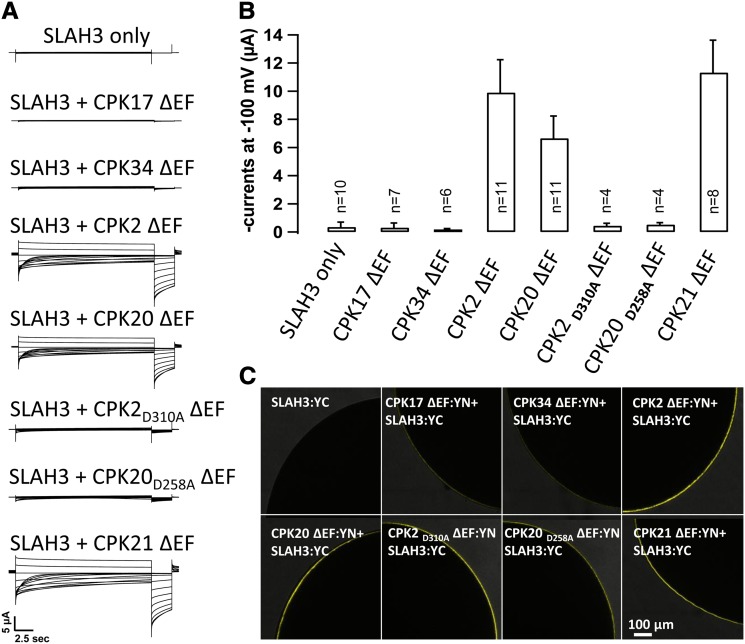
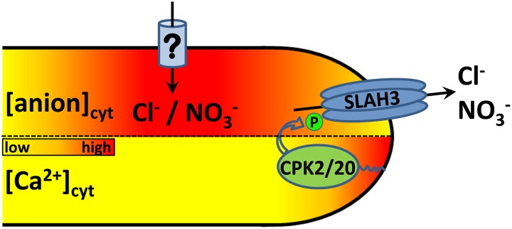
Apical growth in pollen tubes (PTs) is associated with the presence of tip-focused ion gradients and fluxes, implying polar localization or regulation of the underlying transporters. The molecular identity and regulation of anion transporters in PTs is unknown. Here we report a negative gradient of cytosolic anion concentration focused on the tip, in negative correlation with the cytosolic Ca(2+) concentration. We hypothesized that a possible link between these two ions is based on the presence of Ca(2+)-dependent protein kinases (CPKs). We characterized anion channels and CPK transcripts in PTs and analyzed their localization. Yellow fluorescent protein (YFP) tagging of a homolog of SLOW ANION CHANNEL-ASSOCIATED1 (SLAH3:YFP) was widespread along PTs, but, in accordance with the anion efflux, CPK2/CPK20/CPK17/CPK34:YFP fluorescence was strictly localized at the tip plasma membrane. Expression of SLAH3 with either CPK2 or CPK20 (but not CPK17/CPK34) in Xenopus laevis oocytes elicited S-type anion channel currents. Interaction of SLAH3 with CPK2/CPK20 (but not CPK17/CPK34) was confirmed by Förster-resonance energy transfer fluorescence lifetime microscopy in Arabidopsis thaliana mesophyll protoplasts and bimolecular fluorescence complementation in living PTs. Compared with wild-type PTs, slah3-1 and slah3-2 as well as cpk2-1 cpk20-2 PTs had reduced anion currents. Double mutant cpk2-1 cpk20-2 and slah3-1 PTs had reduced extracellular anion fluxes at the tip. Our studies provide evidence for a Ca(2+)-dependent CPK2/CPK20 regulation of the anion channel SLAH3 to regulate PT growth.


Figures

Figure 1.
Figure 1.
Spectroscopic Detection of a Cytosolic Chloride Gradient in Pollen Tubes. Transient (biolistic) expression of Cl--Sensor in N. tabacum pollen tubes. (A) Time-lapse fluorescence false color–coded ratio images of representative pollen tubes subjected to increasing concentrations of chloride (1, 10, 25, and 50 mM Cl, as displayed in the graph). The anion channel inhibitor NPPB was applied in the end of the sequence (see Supplemental Movie 1 online). (B) A kymograph of the same time-series experiments is displayed in the same false colored code. The x-axis and y-axis represent the distance and time, respectively. (C) Magnification of the dotted rectangular selection in (B). Note the especially well-defined oscillations on growth and Cl concentration upon addition of 25 mM Cl (top arrow) and 50 mM Cl (bottom arrow). (D) Simultaneous analysis of growth velocity (red) and cytosolic Cl concentrations in the apical 5 µm (black). Data are from the time series presented in (A) to (C), and are considered to be representative (n = 8). Of special relevance, apical chloride concentration decreases when pollen tube growth accelerates, in an almost perfect counter-phase/negative correlation. [See online article for color version of this figure.]
Figure 2.
Figure 2.
Extracellular Chloride Modifies the Cytosolic Tip-Focused Ca2+ Gradient in Pollen Tubes. Cytosolic Ca2+ concentrations were detected by means of ratiometric measurements of biolistically transformed N. tabacum pollen tubes with the genetically encoded Ca2+ indicator YC3.6. (A) False color–coded kymograph of a representative experiment of a growing pollen tube upon application of 10 mM chloride and 5 µM NPPB (see Supplemental Movie 2 online). (B) Simultaneous analysis of tip-localized [Ca2+free]cyt and growth velocity of the experiment presented in (A). [See online article for color version of this figure.]
Figure 3.
Figure 3.
Identification and Subcellular Localization of the Anion Channel SLAH3 in Pollen Tubes. (A) qRT-PCR analysis of anion channels in pollen tubes. Relative transcript numbers are presented for members of the SLAC/SLAH anion channel family and At ALMT12. RNA extraction and cDNA synthesis were performed on in vitro grown A. thaliana (ecotype Col-0) pollen tubes after 5 h of growth (n = 5). Bar indicates se. (B) SLAH3 promotor-GUS (ProSLAH3::GUS) gene expression in pollen of an A. thaliana flower. A magnification of an anther is presented in the top inlay. A comparison of a pollinated pistil of ProSLAH3::GUS (left) and wild-type Col-0 plant (right) is presented in the bottom right inlay. (C) and (D) Biolistic transformation of N. tabacum pollen with SLAH3:YFP from Arabidopsis demonstrated the uniform plasma membrane localization of the anion channel (D) in contrast with soluble YFP (C). Bar = 10 µm. [See online article for color version of this figure.]
Figure 4.
Figure 4.
Subcellular Localization of All Pollen-Expressed CPKs. Spectroscopic analysis of C-terminal YFP fusion proteins of pollen-expressed CPKs after transient expression in N. tabacum pollen tubes. Images are taken from in vitro grown pollen tubes 5 to 8 h after transformation. Representative fluorescence images are presented from ≥3 independent experiments with numerous pollen tubes recorded. The protein family members are indicated in the figure. Note the plasma membrane localization of CPK2, CPK20, CPK17, and CPK34 at the pollen tube tip. Bar = 10 µm.
Figure 5.
Figure 5.
FRET-FLIM Analysis Identified SLAH3–CPK2/CPK20 Interaction. Average fluorescence lifetime monitored in transiently transformed A. thaliana mesophyll protoplasts with a two-component fit. (A) The average fluorescence lifetime of the donor CFP (mTurquoise) fluorescence is presented when CPKs fused to CFP were expressed alone (open bars) or coexpressed with SLAH3:YFP (filled bars). CPKs coexpressed with SLAH3 are indicated in the figure. The average fluorescence lifetime was compared between CFP and CFP:YFP (fusion protein with a 2 amino acid linker) as control. *P ≤ 0.0002 (paired Student’s t test). (B) Pseudocolor-coded fluorescence lifetime images of an Arabidopsis mesophyll protoplast expressing CPK2:mTurquoise alone (left) or coexpressed with SLAH3:YFP (right). Note the presence of dark blue pixels at the plasma membrane in CPK2:mTurquoise-expressing protoplasts, whereas coexpression with SLAH3:YFP results in fewer dark blue but more cyan and green pixels (chloroplasts are characterized by very short lifetime distributions [red]). [See online article for color version of this figure.]
Figure 6.
Figure 6.
CPK2 and CPK20 Interact with and Activate SLAH3 in X. Laevis Oocytes. (A) Whole-oocyte current recordings upon 20-s voltage pulses ranging from +40 to −180 mV in 20-mV decrements followed by a 3-s voltage pulse to −120 mV. After a preactivation voltage pulse of 0 mV, pronounced instantaneous anion currents could be measured with SLAH3:YC coexpressed with EF hand-truncated versions of CPK2, CPK20, and CPK21, but not with CPK17 or CPK34 fused to the N-terminal half of a YFP (YN). Coexpression of SLAH3:YC with point mutation mutants of CPK2 (CPK2D310A) and CPK20 (CPK20D258A) did not elicit macroscopic anion currents. Of special note, the slow channel deactivation is symbolized by the exponential decrease of negative currents at hyperpolarized membrane potentials. This reflects the weak voltage dependence of the depolarization-activated S-type anion channel SLAH3. (B) The mean instantaneous currents of ≥4 measurements at −100 mV are presented for the coexpression of SLAH3:YC and EF hand-truncated versions of CPK17, CPK34, CPK2, CPK20, CPK2D310A, CPK20D258A, and CPK21 fused to YN. Coexpression of SLAH3:YC with CPK21:YN and only SLAH3:YC (control) were used as positive and negative controls, respectively. *P ≤ 0.0001. Bar indicates sd. (C) Interaction studies by BiFC in the X. laevis oocytes analyzed in (B). Note the YFP fluorescence complementation in oocytes coexpressing CPK2, CPK20, CPK2D310A, CPK20D258A, or CPK21 with SLAH3 but not in those coexpressing CPK17 or CPK34 with SLAH3. [See online article for color version of this figure.]
Figure 7.
Figure 7.
Reduced Anion Currents and Fluxes in slah3-1, slah3-2, and cpk2-1 cpk20-2 Pollen Tubes. (A) Comparison of steady state pollen tube anion currents in NO3-based medium upon application of 400-ms voltage steps from −200 mV to +80 mV (Δ20 mV) when the solution contained 5 mM (○, n = 10), 10 mM (□, n = 10), 20 mM (△, n = 11), or 40 mM NO3 (⋄, n = 10). The inset shows a comparison of the steady state currents in 40 mM Cl-based medium (●, n = 12) or 40 mM NO3-based medium (○, n = 10). (B) Current traces of representative Arabidopsis wild-type Col-0 (top left), slah3-1 (bottom left), slah3-2 (top right), and cpk2-1 cpk20-2 (bottom right) pollen tubes grown in medium containing 40 mM NO3. Note the strongly and moderately reduced anion currents in slah3-1, slah3-2, and cpk2-1 cpk20-2 double mutant pollen tubes. (C) Steady state pollen tube anion currents in NO3-based medium (40 mM) from wild-type Col-0 (○, n = 27), slah3-1 (□, n = 12), slah3-2 (⋄, n = 10), and cpk2-1 cpk20-2 (△, n = 13) pollen tubes. (D) Anion fluxes at the pollen tube tip of wild-type (n = 15, Col-0), cpk2-1 cpk20-2 (n = 15), and slah3-1 (n = 9) pollen tubes measured with anion-selective microelectrodes. (E) Growth rate of the pollen tubes presented in (D). *P ≤ 0.01 (paired Student’s t test). See the Results for statistical analysis of (A) and (C).
Figure 8.
Figure 8.
Model of the Link between Cytosolic Anion and Ca2+ Concentration at the Tip. Tip-localized CPK2 and CPK20 are activated by the high [Ca2+free]cyt at the pollen tube apex. A phosphorylation-dependent activation of SLAH3 by these two kinases takes place exclusively at the pollen tube tip. SLAH3-opening generates a cytosolic concentration gradient with low anion concentrations at the tip. An import of anions at the shank, symbolized by relative high cytosolic anion concentrations, is mediated by yet unknown anion transport proteins. Simplistically, the delays on the oscillatory phases of cytosolic Ca2+ and Cl concentrations could be hypothesized to result from diffusional delays and critical concentration thresholds. This spatial pattern would thus be an emergent property of a double feedback loop of regulation based on the physical properties of these ions. [See online article for color version of this figure.]

Similar articles

Cited by

References

    1. Allen G.J., Chu S.P., Harrington C.L., Schumacher K., Hoffmann T., Tang Y.Y., Grill E., Schroeder J.I. (2001). A defined range of guard cell calcium oscillation parameters encodes stomatal movements. Nature 411: 1053–1057 - PubMed
    1. Allen G.J., Chu S.P., Schumacher K., Shimazaki C.T., Vafeados D., Kemper A., Hawke S.D., Tallman G., Tsien R.Y., Harper J.F., Chory J., Schroeder J.I. (2000). Alteration of stimulus-specific guard cell calcium oscillations and stomatal closing in Arabidopsis det3 mutant. Science 289: 2338–2342 - PubMed
    1. Becker D., Dreyer I., Hoth S., Reid J.D., Busch H., Lehnen M., Palme K., Hedrich R. (1996). Changes in voltage activation, Cs+ sensitivity, and ion permeability in H5 mutants of the plant K+ channel KAT1. Proc. Natl. Acad. Sci. USA 93: 8123–8128 - PMC - PubMed
    1. Becker D., et al. (2004). AtTPK4, an Arabidopsis tandem-pore K+ channel, poised to control the pollen membrane voltage in a pH- and Ca2+-dependent manner. Proc. Natl. Acad. Sci. USA 101: 15621–15626 - PMC - PubMed
    1. Benkert R., Obermeyer G., Bentrup F.-W. (1997). The turgor pressure of growing lily pollen tubes. Protoplasma 198: 1–8

Publication types

MeSH terms

Substances