

Functional analysis of limb recovery following autograft treatment of volumetric muscle loss in the quadriceps femoris
- PMID: 24280565
- PMCID: PMC4017006
- DOI: 10.1016/j.jbiomech.2013.10.057
Functional analysis of limb recovery following autograft treatment of volumetric muscle loss in the quadriceps femoris
Abstract
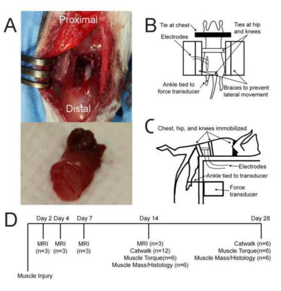
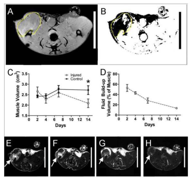
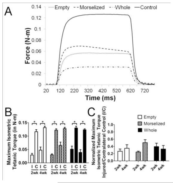
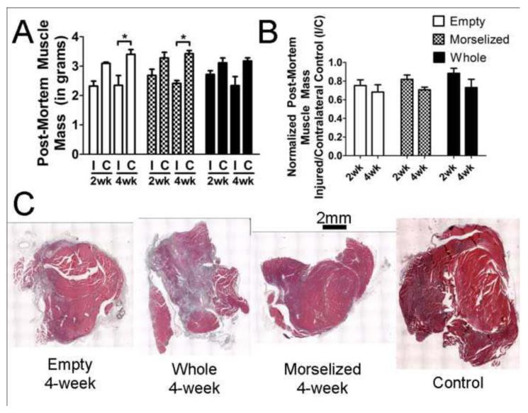
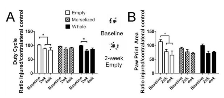
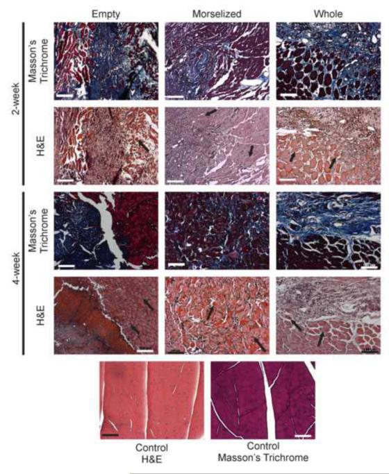
Severe injuries to the extremities often result in muscle trauma and, in some cases, significant volumetric muscle loss (VML). These injuries continue to be challenging to treat, with few available clinical options, a high rate of complications, and often persistent loss of limb function. To facilitate the testing of regenerative strategies for skeletal muscle, we developed a novel quadriceps VML model in the rat, specifically addressing functional recovery of the limb. Our outcome measures included muscle contractility measurements to assess muscle function and gait analysis for evaluation of overall limb function. We also investigated treatment with muscle autografts, whole or minced, to promote regeneration of the defect area. Our defect model resulted in a loss of muscle function, with injured legs generating less than 55% of muscle strength from the contralateral uninjured control legs, even at 4 weeks post-injury. The autograft treatments did not result in significant recovery of muscle function. Measures of static and dynamic gait were significantly decreased in the untreated, empty defect group, indicating a decrease in limb function. Histological sections of the affected muscles showed extensive fibrosis, suggesting that this scarring of the muscle may be in part the cause of the loss of muscle function in this VML model. Taken together, these data are consistent with clinical findings of reduced muscle function in large VML injuries. This new model with quantitative functional outcome measures offers a platform on which to evaluate treatment strategies designed to regenerate muscle tissue volume and restore limb function.
Keywords: Gait analysis; Muscle autograft; Muscle functional testing; Preclinical model; Volumetric muscle loss.
© 2013 Published by Elsevier Ltd.
Figures
References
-
- Bierinx AS, Sebille A. Mouse sectioned muscle regenerates following auto-grafting with muscle fragments: a new muscle precursor cells transfer? Neuroscience letters. 2008;431:211–214. - PubMed
-
- Bozkurt A, Deumens R, Scheffel J, O’Dey DM, Weis J, Joosten EA, Fuhrmann T, Brook GA, Pallua N. CatWalk gait analysis in assessment of functional recovery after sciatic nerve injury. J Neurosci Methods. 2008;173:91–98. - PubMed
-
- Carlson BM, Gutmann E. Development of contractile properties of minced muscle regenerates in the rat. Experimental neurology. 1972;36:239–249. - PubMed
-
- Castillo RC, MacKenzie EJ, Wegener ST, Bosse MJ. Prevalence of chronic pain seven years following limb threatening lower extremity trauma. Pain. 2006;124:321–329. - PubMed
Publication types
MeSH terms
Grants and funding
LinkOut - more resources
Full Text Sources
Other Literature Sources
