. 2014 Feb;10(2):209-25.
doi: 10.4161/auto.26929. Epub 2013 Nov 20.

Role of class III phosphatidylinositol 3-kinase during programmed nuclear death of Tetrahymena thermophila

Affiliations

Role of class III phosphatidylinositol 3-kinase during programmed nuclear death of Tetrahymena thermophila

Takahiko Akematsu et al. Autophagy. 2014 Feb.

Abstract

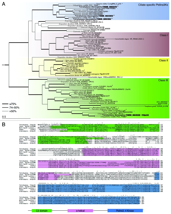
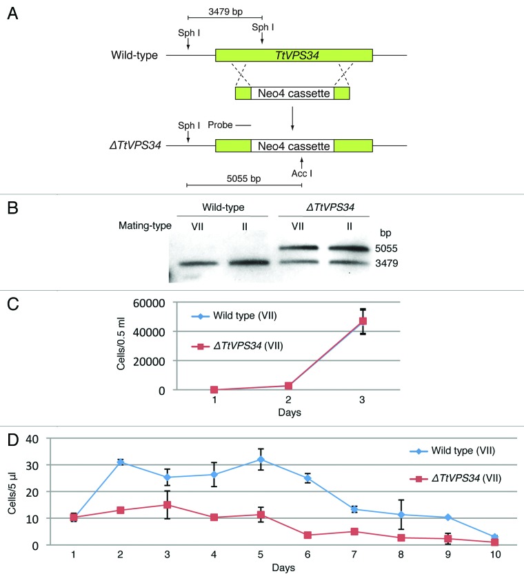
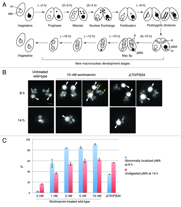
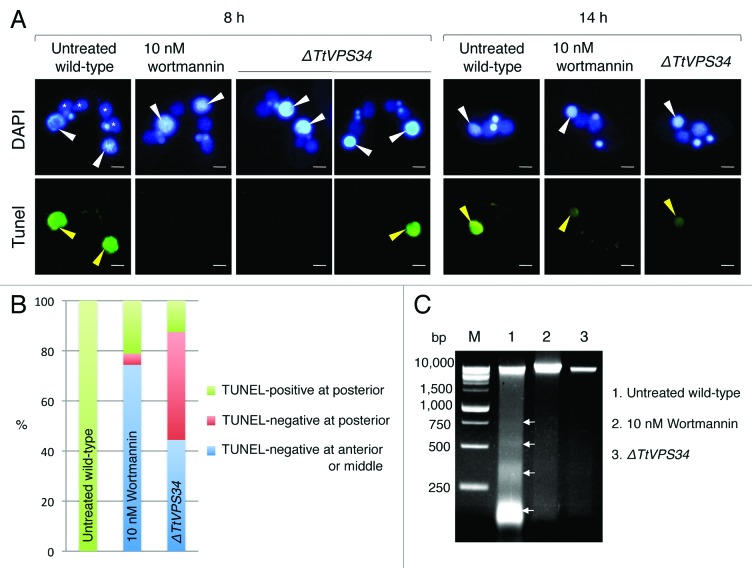
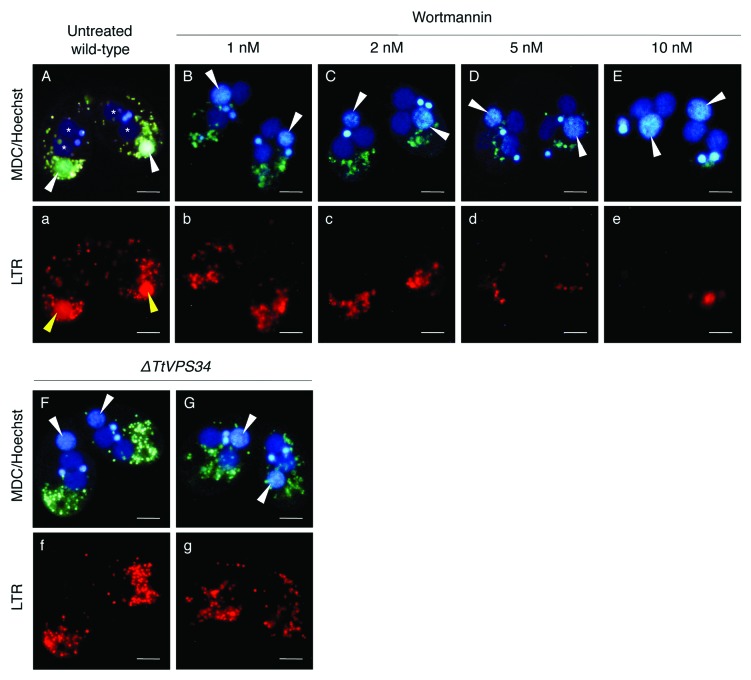
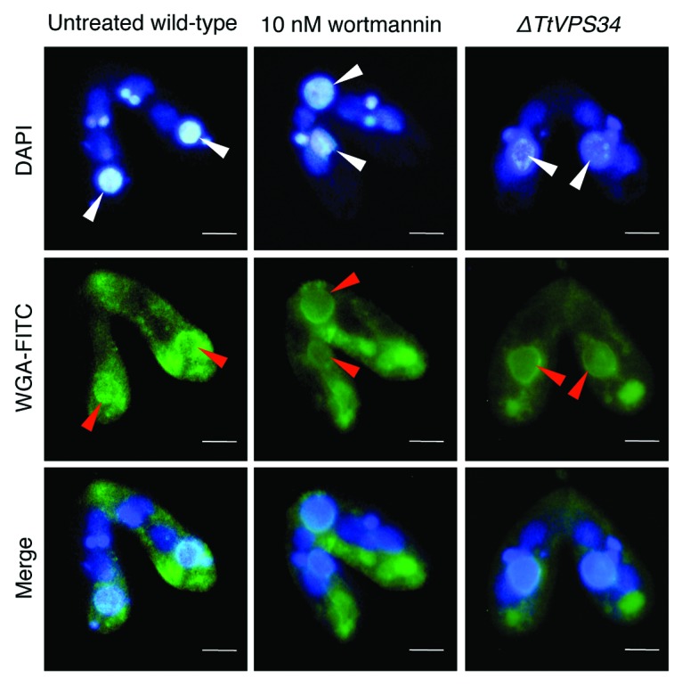
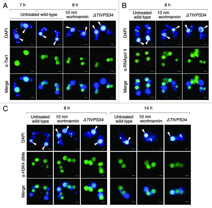
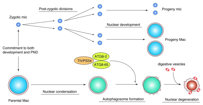
Programmed nuclear death (PND) in the ciliate protozoan Tetrahymena thermophila is a novel type of autophagy that occurs during conjugation, in which only the parental somatic macronucleus is destined to die and is then eliminated from the progeny cytoplasm. Other coexisting nuclei, however, such as new micro- and macronuclei are unaffected. PND starts with condensation in the nucleus followed by apoptotic DNA fragmentation, lysosomal acidification, and final resorption. Because of the peculiarity in the process and the absence of some ATG genes in this organism, the mechanism of PND has remained unclear. In this study, we focus on the role of class III phosphatidylinositol 3-kinase (PtdIns3K, corresponding to yeast Vps34) in order to identify central regulators of PND. We identified the sole Tetrahymena thermophila ortholog (TtVPS34) to yeast Vps34 and human PIK3C3 (the catalytic subunit of PtdIns3K), through phylogenetic analysis, and generated the gene knockdown mutant for functional analysis. Loss of TtVPS34 activity prevents autophagosome formation on the parental macronucleus, and this nucleus escapes from the lysosomal pathway. In turn, DNA fragmentation and final resorption of the nucleus are drastically impaired. These phenotypes are similar to the situation in the ATG8Δ mutants of Tetrahymena, implying an inextricable link between TtVPS34 and TtATG8s in controlling PND as well as general macroautophagy. On the other hand, TtVPS34 does not appear responsible for the nuclear condensation and does not affect the progeny nuclear development. These results demonstrate that TtVPS34 is critically involved in the nuclear degradation events of PND in autophagosome formation rather than with an involvement in commitment to the death program.

Keywords: Atg8; PtdIns3K; Tetrahymena; Vps34; conjugation; macroautophagy; nuclear apoptosis; nuclear development.


Figures

None
Figure 1. Characterization of phosphatidylinositol 3-kinases in Tetrahymena. (A) Phylogenetic tree of PtdIns3Ks. The tree was reconstructed with a maximum likelihood method (see Materials and Methods). Superscripts on the accession numbers stand for databases, which the protein sequences were taken from;†: dictybase, **: Tair, $: Saccharomyces Genome Database, #: WormBase, *:FlyBase, §: GeneDB, ##: OxyDB, ††: ParameciumDB, §§: IchDB, and $$:TGB. The accession numbers without superscript indicate the protein sequences that were taken from GenBank. Branches with different widths represent bootstrap values. Scale bar: 0.5, expected amino acid residue substitutions per site. (B) Multiple-sequence alignment of the whole amino acid sequence of class III PtdIns3Ks (human PIK3C3 or yeast Vps34) including Homo sapiens, Saccharomyces cerevisiae, Dictyostelium discoideum, Arabidopsis thaliana, and Tetrahymena thermophila. Each color box represents the conserved domain, which corresponds to the schematic representation of the primary structure of orthologs of yeast Vps34. Asterisks indicate identical amino acids. Colons and semicolons indicate amino acid similarity.
None
Figure 2. Generation of somatic TtVPS34 gene-knockdown mutants. (A) A schematic showing the TtVPS34 genomic locus (upper), the plasmid vector carrying Neo4 cassette (middle) and after homologous recombination (lower). (B) Southern blot analysis of AccI and SphI digested genomic DNA from wild-type cells and TtVPS34∆ mutants. Molecular weight of the signals against the probe corresponds to the prediction in (A). (C) Cell-growth curves in nutrient-rich conditions. The cells (0.5 ml) maintained in the medium for 1, 2, and 3 d were fixed with paraformaldehyde, diluted 100× and counted under a microscope. (D) Remaining cells in nutrient-deprivation conditions. The cells inoculated in 10 mM TRIS-HCl pH 7.2 were sampled (5 μl) every 24 h until day 10 and swimming (living) cells were counted under a microscope. Points and attached bars correspond to the mean of the 3 identical measurements and standard deviations.
None
Figure 3. Involvement of abnormal localization and lack of digestion of the parental macronucleus with addition of wortmannin or knockdown of TtVPS34. (A) Nuclear events during conjugation of wild-type Tetrahymena. MA, macronucleus; mi, micronucleus; A, progeny macronuclear anlagen; pMA, parental macronucleus; m, progeny micronucleus. (B) Conjugating cells at 8 h (upper) and 14 h (lower) stained with DAPI. White arrowheads, parental macronucleus; asterisks, progeny macronuclear anlagen; dollar signs, progeny micronuclei; yellow arrows, undigested pronuclei. Scale bars: 10 μm. (C) Effects of the treatments on appearance of the abnormalities. Wortmannin was used at a concentration range from 0 to 10 nM. Red and blue columns represent percentage of parental macronucleus (pMA) abnormally localized at 8 h and undigested at 14 h, respectively. The columns and attached bars correspond to the means of 4 identical measurements and standard deviations.
None
Figure 4. A critical role of PtdIns3K activity in DNA degeneration. (A) TUNEL assay results of wortmannin (10 nM)-treated cells and TtVPS34∆ crossings. The cells were sampled at 8 h (left panel) and 14 h (right panel) for the assay. White arrowheads, parental macronucleus; asterisks, developing new macronuclear anlagen; yellow arrowheads, TUNEL-positive parental macronucleus. Scale bars: 10 μm. (B) Relation between reactions of parental macronucleus to TUNEL assay and its localization. Measurements were done on more than 100 cells. (C) Agarose gel electrophoresis using fragmented macronuclear genome extracted from conjugating cells at 8 h. Arrows in the picture correspond to a DNA ladder pattern at ~180 bp intervals. M denotes a DNA sample marker.
None
Figure 5. Effects of wortmannin or knockdown of TtVPS34 on autophagic/lysosomal events during PND. Conjugating cells at 8 h were stained with a combination of MDC (upper) and LTR (lower). Hoechest was also used to visualize nuclei (upper panels). (A, a) Untreated wild-type cross. (B, b–E, e) Wild-type crosses treated with various concentrations of wortmannin ranging from 1 to 10 nM. (F, f) TtVPS34∆ crosses with both the parental macronuclei localized at the anterior region of the cells. (G, g) TtVPS34∆ cross with the parental macronuclei localized at middle (left) and posterior regions of the cytoplasm. White arrowheads, parental macronucleus; asterisks, developing new macronuclear anlagen. Scale bars: 10 μm.
None
Figure 6. Binding of lectin to the parental macronucleus. Conjugating cells at 8 h were fixed and stained with DAPI (upper) and FITC-labeled WGA (middle). The lower parts show a merged image. White arrowheads, parental macronucleus; red arrowheads, concentrated FITC-signal on the nuclear surface. Scale bars: 10 μm.
None
Figure 7. Effects of wortmannin or knockout of TtVPS34 on behavior of Twi1 and hallmarks for active transcription. Conjugating cells were fixed and used for indirect immunofluorescence observations using FITC-labeled antibodies (middle). DAPI was also used to stain nuclei (upper). The lower parts show a merged image. (A) Twi1. (B) Phosphorylated RNA polymerase II at Ser 2 of the C-terminal domain repeat (RNApol-II S2ph). (C) Dimethylation of histone H3 at Lys4 (H3K4 dime). White arrowheads, parental macronucleus; asterisks, developing new macronuclear anlagen. Scale bars: 10 μm.
None
Figure 8. Diagram illustrating a possible role of TtVPS34 in Tetrahymena PND. Once a commitment is made to both development and PND, the zygotic micronucleus and parental macronucleus start to divide and condense, respectively. TtVPS34 plays a critical role in autophagosome formation on the parental macronucleus together with two TtATG8s, which allows digestive vesicles to incorporate with the macronuclear envelope. Both TtVPS34 and TtATG8s do not appear responsible for nuclear condensation and progeny nuclear differentiation.

References

    1. Chalker DL. . Transformation and strain engineering of Tetrahymena. . Methods Cell Biol 2012; 109:327 - 45; http://dx.doi.org/ 10.1016/B978-0-12-385967-9.00011-6; PMID: 22444150 - DOI - PubMed
    1. Coyne RS, Stover NA, Miao W. . Whole genome studies of Tetrahymena. . Methods Cell Biol 2012; 109:53 - 81; http://dx.doi.org/ 10.1016/B978-0-12-385967-9.00004-9; PMID: 22444143 - DOI - PubMed
    1. Orias E, Cervantes MD, Hamilton EP. . Tetrahymena thermophila, a unicellular eukaryote with separate germline and somatic genomes. Res Microbiol 2011; 162:578 - 86; http://dx.doi.org/ 10.1016/j.resmic.2011.05.001; PMID: 21624459 - DOI - PMC - PubMed
    1. Iwamoto M, Mori C, Kojidani T, Bunai F, Hori T, Fukagawa T, Hiraoka Y, Haraguchi T. . Two distinct repeat sequences of Nup98 nucleoporins characterize dual nuclei in the binucleated ciliate tetrahymena. . Curr Biol 2009; 19:843 - 7; http://dx.doi.org/ 10.1016/j.cub.2009.03.055; PMID: 19375312 - DOI - PubMed
    1. Cole E, Sugai T. . Developmental progression of Tetrahymena through the cell cycle and conjugation. Methods Cell Biol 2012; 109:177 - 236; http://dx.doi.org/ 10.1016/B978-0-12-385967-9.00007-4; PMID: 22444146 - DOI - PubMed

Publication types

MeSH terms

Substances