. 2014 Jan 31;114(3):480-92.
doi: 10.1161/CIRCRESAHA.114.302113. Epub 2013 Nov 26.

Activation of NAD(P)H oxidase by tryptophan-derived 3-hydroxykynurenine accelerates endothelial apoptosis and dysfunction in vivo

Affiliations

Activation of NAD(P)H oxidase by tryptophan-derived 3-hydroxykynurenine accelerates endothelial apoptosis and dysfunction in vivo

Qiongxin Wang et al. Circ Res. .

Abstract

Rationale: The kynurenine (Kyn) pathway is the major route for tryptophan (Trp) metabolism in mammals. The Trp-Kyn pathway is reported to regulate several fundamental biological processes, including cell death.

Objective: The aim of this study was to elucidate the contributions and molecular mechanism of Trp-Kyn pathway to endothelial cell death.

Methods and results: Endogenous reactive oxygen species, endothelial cell apoptosis, and endothelium-dependent and endothelium-independent vasorelaxation were measured in aortas of wild-type mice or mice deficient for nicotinamide adenine dinucleotide phosphate [NAD(P)H] oxidase subunits (p47(phox) or gp91(phox)) or indoleamine-pyrrole 2,3-dioxygenase 1 with or without angiotensin (Ang) II infusion. As expected, AngII increased plasma levels of Kyn- and 3-hydroxykynurenine-modified proteins in endothelial cells in vivo. Consistent with this, AngII markedly increased the expression of indoleamine-pyrrole 2,3-dioxygenase in parallel with increased expression of interferon-γ. Furthermore, in wild-type mice, AngII significantly increased oxidative stress, endothelial cell apoptosis, and endothelial dysfunction. These effects of AngII infusion were significantly suppressed in mice deficient for p47(phox), gp91(phox), or indoleamine-pyrrole 2,3-dioxygenase 1, suggesting that AngII-induced enhancement of Kynurenines via NAD(P)H oxidase-derived oxidants causes endothelial cell apoptosis and dysfunction in vivo. Furthermore, interferon-γ neutralization eliminates AngII-increased superoxide products and endothelial apoptosis by inhibiting AngII-induced Kynurenines generation, suggesting that AngII-activated Kyn pathway is interferon-γ-dependent. Mechanistically, we found that AngII-enhanced 3-hydroxykynurenine promoted the generation of NAD(P)H oxidase-mediated superoxide anions by increasing the translocation and membrane assembly of NAD(P)H oxidase subunits in endothelial cells, resulting in accelerated apoptosis and consequent endothelial dysfunction.

Conclusions: Kyn pathway activation accelerates apoptosis and dysfunction of the endothelium by upregulating NAD(P)H-derived superoxide.

Keywords: 3-hydroxykynurenine; NAD(P)H oxidase; apoptosis; indoleamine-pyrrole 2,3,-dioxygenase; kynurenine; oxidative stress; tryptophan.


Figures

Figure 1
Figure 1. P47phox or gp91phox deletion attenuates AngII infusion-mediated endothelial apoptosis and dysfunction, in vivo
WT, P47phox−/− and gp91phox−/− mice were infused with saline or AngII (1000ng/min*Kg) for 2 weeks. A and B, Deletion of p47phox and gp91phox ablates AngII-induced superoxide production in vivo. AngII-stimulated superoxide anions were detected by using DHE staining, as described in Materials and Methods. C, Deletion of p47phox and gp91phox abolishes AngII-induced endothelial cell apoptosis. Apoptotic cells were detected by TUNEL-positive cells in ECs (n=4–6 per group, n=4 sections for each sample). D and E, p47phox or gp91phox deficiency rescues endothelial dysfunction induced by AngII. Endothelial function was assayed by Ach induced endothelium-dependent relaxation in WT, p47phox−/− and gp91phox−/− mice aortas. F and G, SNP induced endothelium-independent relaxation in WT, p47phox−/− and gp91phox−/− mice aortas are not affected by AngII. *, p<0.05 versus WT-saline; **, p<0.01 versus WT-saline; #, p<0.05 versus WT- AngII, n=4 to 10 per group.
Figure 2
Figure 2. IDO deletion alleviates endothelial apoptosis and dysfunction via superoxide anions elimination, in vivo
WT and IDO−/− mice were administrated with saline or AngII (1000ng/min*kg) for 4 weeks. A, AngII increases Kyn. The serum concentrations of Kyn were detected by HPLC. B, AngII increases IDO expression in vivo. C, AngII increases the detection of 3-OHkyn modified proteins in WT but not in IDO−/− mice. B and C, Representative immunohistochemical staining (original magnification, X400) and quantifications for IDO (B) and 3-OHkyn modified proteins (C) in aortas. D, AngII increases superoxide anions in WT but not in IDO−/− mice. Superoxide anions were detected by using DHE staining. E and F, IDO deletion abrogates AngII-mediated endothelial apoptosis. Triple immunofluorescence photographs show apoptotic ECs (green) in WT and IDO−/− mice aortic endothelium (CD 31, red); nuclei (Hoechst, blue). TUNEL positive cells colocalized with CD31 positive cells and CD31 positive cells were counted in all aortic sections (original magnification, 25×). Histogram shows the mean number of TUNEL-CD31 positive nuclei/CD31 positive nuclei. Images are representatives of a study conducted in a separate set of animals (n=6 per group, n=4 sections for each sample). G and H, The endothelium-dependent relaxation and -independent relaxation in mouse aortas of WT and IDO−/− with or without AngII. G, IDO deficiency improves the impairment of endothelial function caused by AngII. H, SNP induced endothelium-independent relaxation in mice aortas of WT and IDO−/− are not affected by AngII. *, p<0.01 versus WT-saline; #, p<0.05 versus WT-AngII, n=4 to 10 per group.
Figure 3
Figure 3. Both AngII-increased levels of Kyn and 3-OHkyn-modified proteins and AngII-induced superoxide anions and endothelial apoptosis are IFN-γ-mediated in mouse aortas in vivo
A, WT and IDO−/− mice were administrated with saline or AngII (1000ng/min*kg) for 4 weeks. AngII increases the detection of IFN-γ in both of WT and IDO−/− mice. B–F, WT mice were intraperitoneally injected with anti-IFN-γ or control rat IgG1 antibodies for 4 weeks beginning on the day of AngII infusion. B, IFN-γ neutralization eliminates Kyn. The serum concentrations of Kyn were detected by HPLC. C, IFN-γ neutralization inhibits IDO expression. D, IFN-γ neutralization ameliorates 3-OHkyn modified protein levels. E, IFN-γ neutralization abrogates AngII-induced superoxide anions in vivo. F, IFN-γ neutralization abolishes AngII-induced active caspase-3 staining. A, C, D and F, Representative immunohistochemical staining (original magnification, X400) and quantifications for the levels of IFN-γ (A), IDO (C), 3-OHkyn modified proteins (D) and active caspase-3 (F) in aortas, WT mice infused with saline for 4 weeks were employed as control (C, D and F). *, p<0.01 versus WT saline or WT AngII-anti-IgG, n=4 to 6 per group.
Figure 4
Figure 4. The inhibition of IDO or KMO abrogates ROS generation and cell apoptosis induced by IFN-γ
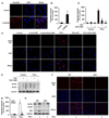
A, IFN-γ-induced endothelial cell apoptosis is dose-dependent. HAECs were incubated for 48 hours with various concentration of IFN-γ. B, IFN-γ-induced endothelial cell apoptosis is time-dependent. HAECs were treated with 100ng/ml IFN-γ for different time. C–F, HAECs were pretreated for 1 hour with medium alone or medium containing MT (200µM) or Ro61-8048 (50 µM) before incubation with or without IFN-γ (100ng/ml) for 48 hours. G–J, MLECs isolated from WT and IDO−/− mice were challenged with or without 100ng/ml IFN-γ for 48 hours. C and G, Pretreatment with IDO inhibitor (1-MT) or KMO inhibitor (Ro61-8048) (C), or IDO deficiency (G) inhibits IFN-γ-induced protein cleavage of parp, caspase-3 and caspase-7 in ECs in vitro. D and H, Inhibition of IDO (D & H) or KMO (D) blocks IFN-γ-induced caspase-3 activation in ECs. E & I, Inhibition of IDO (E & I) or KMO (E) eliminates IFN-γ-induced cell apoptosis. F & J, Inhibition of IDO (F and J) or KMO (F) attenuates IFN-γ-induced ROS formation in ECs. A–C and G, Cell lysates were subjected to immunoblot analysis to detect the protein cleavage of PARP, caspase-7 and caspase-3. GAPDH and β-actin were used as loading controls. D and H, Capase-3 activity was measured in a fluorescent microplate reader. The results were the mean ± S.E. of three independent measurements. E and I, Nuclear DNA fragment as a sign of apoptosis was detected using a TUNEL assay. Data were reported as %TUNEL-positive in a microscopic field ±S.E. F and J, Superoxide production was assessed by DHE/HPLC. *, p<0.05 versus control; **, p<0.01 versus control; #, p<0.05 versus IFN- γ (A–F) or WT IFN-γ (G–J); ##, p<0.01 versus IFN- γ (A–F) or WT IFN-γ (G–J).
Figure 5
Figure 5. IFN-γ induces IDO and KMO protein expressions and endogenous kynurenine formation
A–C, HAECs were incubated for 24 hours with various concentration of IFN-γ. D–F, HAECs were treated with 100ng/ml IFN-γ for different time. A and D, IFN-γ induces the expressions of IDO and KMO in a dose- and time- dependent manner. Cell lysates were subjected to immunoblot analysis to detect IDO, KMO and β-actin. B and E, IFN-γ increases Kyn but decreases Trp in a dose- and time- dependent manner. C and F, IFN-γ increases IDO activity in a dose- and time- dependent manner. G–I, Inhibition of IDO reduces IFN-γ induced Kyn formation and Trp consumption. G, HAECs were pretreated for 1 hour with medium alone or medium containing 200µM 1-MT before incubation with or without IFN-γ (100ng/ml) for 48 hours. H and I, MLECs were stimulated with or without 100ng/ml IFN-γ for 48 hours. B, E and G–I, The levels of Trp and kyn in the culture medium were determined by HPLC. *, p<0.05 versus control; **, p<0.01 versus control; #, p<0.05 versus IFN-γ (G) or WT IFN-γ (H & J); ##, p<0.01 versus WT IFN-γ.
Figure 6
Figure 6. IFN-γ induced 3-OHkyn is both IDO- and KMO-dependent
A and B, Exogenous 3-OHkyn reacts with intracellular proteins. Haecs were incubated with or without 60µM 3-OHkyn for 24 hours. C–H, Inhibition of IDO or KMO (C–E), or IDO deficiency (F–H) eliminates 3-OHkyn modification of intracellular proteins induced by IFN-γ. C–E, Haecs were pretreated for 1 hour with medium alone or medium containing 200µM 1-MT or 50 µM Ro61-8048 before incubation with or without 100ng/ml IFN-γ for 48 hours. F–H, MLECs were challenged with or without 100ng/ml IFN-γ for 48 hours. A, C and F, Immunocytochemistry was performed to localize 3-OHkyn modifications by mouse anti-3-OHkyn monoclonal primary antibody and Alexa594 red-conjugated goat anti-mouse IgG secondary antibody. Cells were counterstained with a nuclear stain, DAPI (blue) (From up: 3-OHkyn modifications, DAPI and merge). E and H, Cell lysates were subjected to immunoblot analysis to detect 3-OHkyn modified proteins. GAPDH and β-actin were used as loading controls. *, p<0.01 versus control; #, p<0.01 versus IFN- γ.
Figure 7
Figure 7. 3-OHkyn is apoptosis-inducing kynurenine
A–C, 3-OHkyn induces cell apoptosis. HAECs were treated with complete serum medium or serum free medium alone or containing Kyn (60µM) or 3-OHkyn for 24 hours. D–F, 3-OHkyn induces cell apoptosis in a dose-dependent manner. HAECs were incubated for 24 hours with various concentration of 3-OHkyn. G–I, 3-OHkyn induces cell apoptosis in a time-dependent manner. HAECs were treated with 60µM 3-OHkyn for different time. A, D and G, Cell lysates were subjected to immunoblot analysis to detect the protein cleavage of parp, caspase-7 and caspase-3. B, E and H, The results of capase-3 activity were the mean ± S.E. of three independent measurements. C, F and I, Data were reported as %TUNEL-positive in a microscopic field ±S.E. *, p<0.05 versus SFM or control; **, p<0.01 versus control.
Figure 8
Figure 8. 3-OHkyn reduces cell viability but induces mitochondrial cytochrome c release, and ROS formation in a NAD(P)H oxidase-dependent manner
A–C, HAECs were incubated for 24 hours with indicated concentration of 3-OHkyn. D–J, HAECs were incubated with or without 3-OHkyn (60 µM) for 24 hours, which were infected with ad-GFP or ad-SOD1 (G)/2 (D), or pretreated with medium alone or medium containing tempol (10µM) or apocynin (100µM) for 1 hour (E & F). A, 3-OHkyn reduces endothelial cell viability in a dose-dependent manner. Cell viability was assessed by using MTT reduction assays. B and E, 3-OHkyn induces intracellular ROS generation, which is ablated by NAD(P)H oxidase inhibitor. Superoxide production was assessed by DHE/HPLC. C, 3-OHkyn accelerates mitochondrial cytochrome c release. Cytochrome c was detected in cytosolic fractions using a monoclonal antibody. D, F and G, Overexpress of SOD or pretreatment with NAD(P)H oxidase inhibitor attenuates 3-OHkyn-triggered apoptosis. Cell lysates were subjected to immunoblot analysis to detect SOD1 (G) or 2 (D) and the protein cleavage of PARP, caspase-7 and caspase-3. H, 3-OHkyn promotes translocation and membrane assembly of p47phox and p67phox. Immunoblot analysis of NAD(P)H oxidase subunits (p47phox and p67phox) were applied in membrane proteins and whole cell lysates by immunoblot. I, 3-OHkyn increases NAD(P)H activity. NAD(P)H activity was measured by a low concentration based lucigenin (5µM) assay. The results were the mean ± S.E. of three independent measurements. J, 3-OHkyn reacts with and chemically modifies p47phox and p67phox. Cell lysates were subjected to immunoprecipitation (IP)/immunoblot analysis to detect p47phox and p67phox modified by 3-OHkyn. Na+/K+ ATPase and β-actin were used as loading controls. *, p<0.05 versus control; **, p<0.01 versus control; #, p<0.05 versus 3-OHkyn only.

Comment in

  • Do endothelial cells eat tryptophan to die?
    Duran C, San Martín A. Duran C, et al. Circ Res. 2014 Jan 31;114(3):406-8. doi: 10.1161/CIRCRESAHA.113.303150. Circ Res. 2014. PMID: 24481839 Free PMC article. No abstract available.

References

    1. Stefanec T. Endothelial apoptosis: Could it have a role in the pathogenesis and treatment of disease? Chest. 2000;117:841–854. - PubMed
    1. Galley HF, Webster NR. Physiology of the endothelium. British journal of anaesthesia. 2004;93:105–113. - PubMed
    1. Cai H, Harrison DG. Endothelial dysfunction in cardiovascular diseases: The role of oxidant stress. Circulation research. 2000;87:840–844. - PubMed
    1. Sprague AH, Khalil RA. Inflammatory cytokines in vascular dysfunction and vascular disease. Biochemical pharmacology. 2009;78:539–552. - PMC - PubMed
    1. King NJ, Thomas SR. Molecules in focus: Indoleamine 2,3-dioxygenase. The international journal of biochemistry & cell biology. 2007;39:2167–2172. - PubMed

Publication types