

Circle of Willis configuration as a determinant of intracranial dolichoectasia
- PMID: 24281350
- PMCID: PMC4370622
- DOI: 10.1159/000356347
Circle of Willis configuration as a determinant of intracranial dolichoectasia
Abstract
Background: Circle of Willis (COW) variants might influence arterial caliber in the brain. We hypothesized that these variants would be associated with the prevalence of intracranial dolichoectasia (DE).
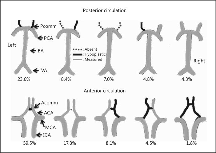
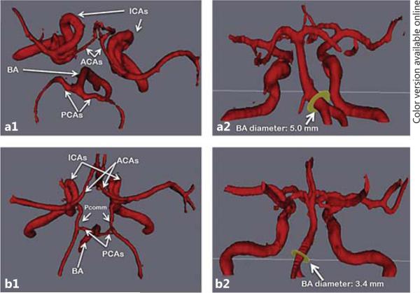
Methods: We examined COW variants and DE in a sample of stroke-free participants (n = 436) undergoing magnetic resonance angiography (MRA) as part of a population-based study. Large intracranial arterial diameters were obtained when available; if not, the artery was defined as hypoplastic or absent according to its visibility on MRA. Subscores for the anterior and the posterior circulations were created. DE was defined as arterial diameters ≥2 SD above the population mean for that artery, adjusting for intracranial volume. Generalized linear models with a Poisson distribution were used to evaluate predictors of both absent and hypoplastic vessels, and logistic regression was used to assess the odds ratio (OR) and 95% confidence interval (95% CI) of DE depending on COW variants.
Results: Only 44% of the sample had all 14 arteries present, 32% lacked 1 artery, 18% lacked 2 and 6% lacked 3 or more. DE of at least 1 artery was not associated with the total number of hypoplastic or absent arteries, but DE in a posterior circulation artery was weakly associated with the number of absent arteries in the posterior circulation (β coefficient = 0.36, p = 0.06). DE of at least 1 artery was more frequent in those with 1 or more absent arteries (OR 1.27, 95% CI 1.03-1.57). Posterior circulation DE was more frequent in participants with at least 1 or more absent arteries at any location (OR 1.35, 95% CI 1.02-1.78). Participants with an incomplete posterior COW were more likely to have DE in the anterior circulation (OR 1.52, 95% CI 1.01-2.33). Having an absent left anterior cerebral artery (ACA) A1 segment was associated with right ACA DE (OR 34.1, 95% CI 3.16-368.2); an absent right ACA was associated with left ACA DE (OR 14.1, 95% CI 1.69-118.28). Absence of 1 (OR 1.9, 95% CI 1.1-3.4) or 2 (OR 3.0, 95% CI 1.4-6.6) of the 2 arteries connecting the anterior to the posterior circulation was associated with basilar artery DE.
Conclusion: The COW is a pleomorphic structure that allows collateral flow to compensate for an insufficient or absent arterial component at the base of the skull. By presumed flow diversion, arteries might undergo outward remodeling. Whether this compensatory arterial dilatation is beneficial or not remains unknown.
Figures

Similar articles
-
Incomplete circle of Willis is associated with a higher incidence of neurologic events during carotid eversion endarterectomy without shunting.J Vasc Surg. 2018 Dec;68(6):1764-1771. doi: 10.1016/j.jvs.2018.03.429. Epub 2018 Jul 6. J Vasc Surg. 2018. PMID: 29983353
-
Correlation between Circle of Willis configuration and intracranial arterial dolichoectasia, and genetic contributions.J Stroke Cerebrovasc Dis. 2024 Nov;33(11):107955. doi: 10.1016/j.jstrokecerebrovasdis.2024.107955. Epub 2024 Aug 22. J Stroke Cerebrovasc Dis. 2024. PMID: 39179190
-
Takayasu arteritis: ultrasonographic evaluation of the cervico-cerebral arteries.Int J Cardiol. 1998 Oct 1;66 Suppl 1:S163-73. doi: 10.1016/s0167-5273(98)00164-8. Int J Cardiol. 1998. PMID: 9951816
-
The fetal variant of the circle of Willis and its influence on the cerebral collateral circulation.Cerebrovasc Dis. 2006;22(4):217-24. doi: 10.1159/000094007. Epub 2006 Jun 20. Cerebrovasc Dis. 2006. PMID: 16788293 Review.
-
Morphological aspects of the vasculogenesis and angiogenesis during prenatal edification of the circle of Willis: a review.Rom J Morphol Embryol. 2021 Jul-Sep;62(3):679-687. doi: 10.47162/RJME.62.3.04. Rom J Morphol Embryol. 2021. PMID: 35263395 Free PMC article. Review.
Cited by
-
A 5-Year Follow-up of Intracranial Arterial Dolichoectasia: A Case Report and Review of Literature.Asian J Neurosurg. 2019 Nov 25;14(4):1302-1307. doi: 10.4103/ajns.AJNS_282_18. eCollection 2019 Oct-Dec. Asian J Neurosurg. 2019. PMID: 31903384 Free PMC article.
-
A quantitative perspective to the study of brain arterial remodeling of donors with and without HIV in the Brain Arterial Remodeling Study (BARS).Front Physiol. 2014 Feb 19;5:56. doi: 10.3389/fphys.2014.00056. eCollection 2014. Front Physiol. 2014. PMID: 24600402 Free PMC article.
-
Association of brain arterial diameters with demographic and anatomical factors in a multi-national pooled analysis of cohort studies.Neuroradiol J. 2024 Jun;37(3):304-313. doi: 10.1177/19714009231224429. Epub 2023 Dec 26. Neuroradiol J. 2024. PMID: 38148489 Free PMC article.
-
Vascular variants and the evaluation of patients with acute stroke.Neurol Clin Pract. 2019 Jun;9(3):185-186. doi: 10.1212/CPJ.0000000000000635. Neurol Clin Pract. 2019. PMID: 31342959 Free PMC article. No abstract available.
-
Anatomical effects on the relationship between brain arterial diameter and length: The Northern Manhattan Study.J Neuroimaging. 2022 Jul;32(4):735-743. doi: 10.1111/jon.12969. Epub 2022 Jan 26. J Neuroimaging. 2022. PMID: 35083811 Free PMC article.
References
-
- Passero SG, Calchetti B, Bartalini S. Intracranial bleeding in patients with vertebrobasilar dolichoectasia. Stroke. 2005;36:1421–1425. - PubMed
-
- Flemming KD, Wiebers DO, Brown RD, Jr, Link MJ, Huston J, 3rd, McClelland RL, Christianson TJ. The natural history of radiographically defined vertebrobasilar nonsaccular intracranial aneurysms. Cerebrovasc Dis. 2005;20:270–279. - PubMed
-
- Ubogu EE, Chase CM, Verrees MA, Metzger AK, Zaidat OO. Cervicomedullary junction compression caused by vertebral artery dolichoectasia and requiring surgical treatment. Case report. J Neurosurg. 2002;96:140–143. - PubMed
MeSH terms
Grants and funding
LinkOut - more resources
Full Text Sources
Other Literature Sources
