. 2014 May;133(5):599-616.
doi: 10.1007/s00439-013-1391-3. Epub 2013 Nov 27.

COOH-terminal collagen Q (COLQ) mutants causing human deficiency of endplate acetylcholinesterase impair the interaction of ColQ with proteins of the basal lamina

Affiliations

COOH-terminal collagen Q (COLQ) mutants causing human deficiency of endplate acetylcholinesterase impair the interaction of ColQ with proteins of the basal lamina

Juan Arredondo et al. Hum Genet. 2014 May.

Abstract

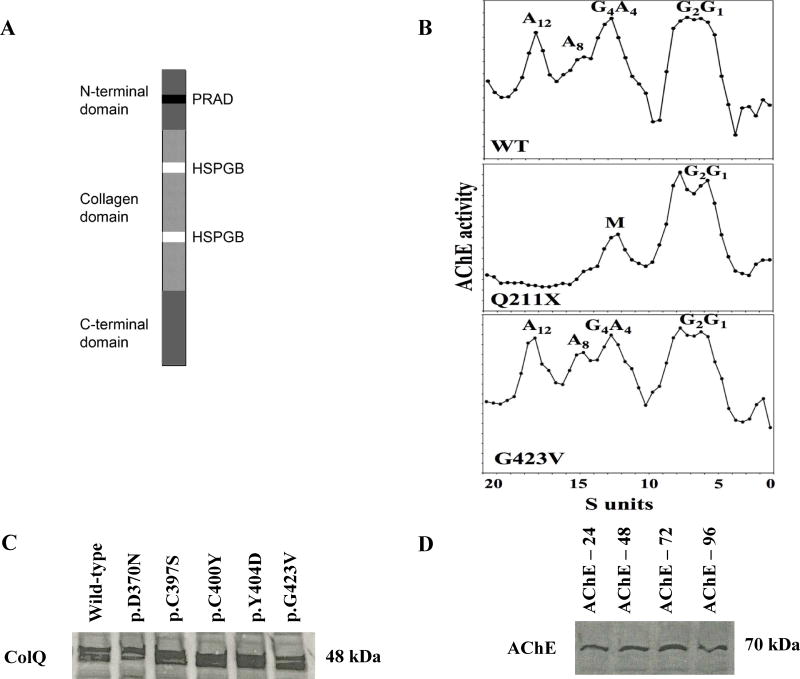
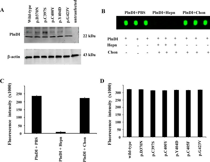
Collagen Q (ColQ) is a key multidomain functional protein of the neuromuscular junction (NMJ), crucial for anchoring acetylcholinesterase (AChE) to the basal lamina (BL) and accumulating AChE at the NMJ. The attachment of AChE to the BL is primarily accomplished by the binding of the ColQ collagen domain to the heparan sulfate proteoglycan perlecan and the COOH-terminus to the muscle-specific receptor tyrosine kinase (MuSK), which in turn plays a fundamental role in the development and maintenance of the NMJ. Yet, the precise mechanism by which ColQ anchors AChE at the NMJ remains unknown. We identified five novel mutations at the COOH-terminus of ColQ in seven patients from five families affected with endplate (EP) AChE deficiency. We found that the mutations do not affect the assembly of ColQ with AChE to form asymmetric forms of AChE or impair the interaction of ColQ with perlecan. By contrast, all mutations impair in varied degree the interaction of ColQ with MuSK as well as basement membrane extract (BME) that have no detectable MuSK. Our data confirm that the interaction of ColQ to perlecan and MuSK is crucial for anchoring AChE to the NMJ. In addition, the identified COOH-terminal mutants not only reduce the interaction of ColQ with MuSK, but also diminish the interaction of ColQ with BME. These findings suggest that the impaired attachment of COOH-terminal mutants causing EP AChE deficiency is in part independent of MuSK, and that the COOH-terminus of ColQ may interact with other proteins at the BL.


Figures

Fig. 1
Fig. 1
The COOH-terminal ColQ mutations do not impair assembly of AChE with ColQ. (a) Schematic diagram showing the central triple-helical collagen domain is surrounded by non-collagenous NH2-terminal and COOH-terminal domains of ColQ. The PRAD region is within the NH2-terminal domain and the two HSPG-binding (HSPGB) motifs are located in the collagen domain. (b) Representative sucrose density gradients. Truncation mutations such as p. Gln211* result only in the globular forms of the enzyme (G1 and G2) plus an M peak. COOH-terminal mutations such as p.Gly423Val do not alter the assembly of ColQ with AChET and the sedimentation profile is similar to that of WT. The amplitudes of the A12 and the G1 and G2 peaks are highly variable from preparation to preparation. (c) Overexpression of ColQ protein on COS-7 cells transfected with WT ColQ and mutant constructs. Crude cell lysates were immunoblotted with ColQ antibody. (d) Crude cell lysates of AChE overexpressed protein on COS-7 cells were immunoblotted with AChE antibody. Western blot variations of transfection efficiency were corrected by monitoring the expression of β-actin in the immunoblots, data not shown.
Fig. 2
Fig. 2
The COOH-terminal ColQ mutations do not impair binding of ColQ subunit to perlecan. (a) Co-immunoprecipitation experiments were performed after co-expression of AChET and WT ColQ or mutants in inducible HEK-EBNA cells expressing PlnDI. The protein complex between ColQ and perlecan proteins was co-immunoprecipitated with goat antibody against ColQ. The immunoprecipitates were digested with heparitinases (50 IU/g protein) and chondroitinase (1 U/mg protein), and analyzed by Western blots using rabbit antibody against perlecan. The housekeeping β-actin protein was used to normalize the protein expression. Only the control lane, non-transfected HEK-EBNA cells, showed no PlnDI band, ~22 kDa. Endogenous perlecan will not appear in the blots due to its enormous size and glycosaminoglycan chains. (b) Dot-blot analyses of ColQ interaction with PlnDI. The PlnDI was immobilized on nitrocellulose in its native form or after digestion with heparinases I, II, and III (Hepn) or chondroitinase AC (Chon). A12 form binding was determined by ColQ antibody, the samples were blotted in triplicate on nitrocellulose membrane. (c) Bar graph summarizes fluorescence measurements performed on dot blot. (d) Summary of the dot blots binding assays. The purified AChE A12 asymmetric forms for each mutant were incubated on nitrocellulose membrane blotted with PlnDI. The bar graph summarizes measurements dot blot performed on each mutant.
Fig. 3
Fig. 3
The COOH-terminal ColQ mutations impair the binding of the ColQ subunit to MuSK. (a) Co-immunoprecipitation experiments were performed after co-transfection of HEK-293 cells with either WT ColQ or one of the ColQ mutants, p.Asp370Asn, p.Cys397Ser, p.Cys400Tyr, p.Tyr404Asp, or Gly423Val, and with WT MuSK. The protein complex between MuSK and ColQ proteins was co-immunoprecipitated with rabbit MuSK antibody. The immunoprecipitates were analyzed by Western blots using goat antibody against ColQ. The WT ColQ protein (48 kDa) showed the strongest interaction with MuSK, while p.Gly423Val showed the weakest interaction with MuSK. The control lane, un-transfected HEK cells, showed no ColQ band. The housekeeping β-actin protein was used as a control to verify that all the samples had the same amount of loaded protein. (b) The binding assays, where the 96-well plate was coated with pure MuSK. The purified AChE A12 asymmetric forms were overlaid on the plate. The AChE enzymatic activity was used to measure the binding of AChE A12 asymmetric forms to MuSK. Only results for the mutants p.Cys400Tyr, p.Tyr404Asp, p.Cys405Phe and p.Gly423Val had statistical significance (*P < 0.05).
Fig. 4
Fig. 4
The COOH-terminal ColQ mutations impair binding of ColQ subunit to the basal lamina. (a) The binding assays, where the 96-well plate was coated with BME. The purified AChE A12 asymmetric forms from each mutant and wild type were overlaid on top of BME. The binding affinity of ColQ was measured by In-Cell Western, where the ColQ binding was measured by the fluorescence intensity. In comparison with the WT, all mutants showed different levels of fluorescence intensity. The globular AChE forms were used as a negative control. (b) The quantitative analysis shown in the graph demonstrates that the WT had the highest fluorescent signal intensity; mutants p.Asp370Asn, and p.Cys397Ser did not have statistical significance, and p.Cys400Tyr, p.Tyr404Asp, p.Cys405Phe and p.Gly423Val had the lowest fluorescent intensity, all statistically significant (*P < 0.05).
Fig. 5
Fig. 5
The COOH-terminal ColQ mutations do not affect AChE enzymatic activity. Purified A12 asymmetric forms were used to quantify the AChE enzymatic activity in solution. The WT and p.Asp370Asn, p.Cys397Ser, p.Cys400Tyr, p.Tyr404Asp, p.Cys405Phe, and p.Gly423Val mutants purified A12 forms’ AChE activity was quantified by Ellman assay. The results confirmed that COOH-terminal ColQ mutations did not affect the enzyme efficiency. The graph showed a linear trend over time. The data showed that AChE tetramers attached to all ColQ mutants had the same AChE esterase activity as the WT ColQ subunit attached to the AChE catalytic subunit.
Fig. 6
Fig. 6
The COOH-terminal ColQ mutations impair binding of asymmetric AChE forms to BME. The purified A12 AChE asymmetric forms were incubated with BME. Then, the AChE esterase enzymatic activity was measured at different time points as shown in the graph by performing the Ellman assay. The data showed that mutants’ p.Asp370Asn and p.Cys397Ser had slightly lower levels of AChE esterase activity than WT. The mutants p.Cys400Tyr, p.Tyr404Asp, and p.Cys405Phe had lower enzymatic activity than WT, and the p.Gly423Val showed the lowest level of AChE enzymatic activity; all were statistically significant (*P < 0.05). The globular AChE forms were used as a negative control.
Fig. 7
Fig. 7
Theoretical tertiary structure of COOH-terminal ColQ subunit (single strand showing only). The 3D structure is represented in ribbon diagram; the α-helices are in magenta, β-sheets in yellow, and coil in green. The mutated residue side chains are illustrated as follows: p.D370N in blue, p.Cys397Ser in pink, p.Cys400Tyr in yellow, p.Tyr404Asp in red, p.Cys405Phe in green, and p.Gly423Val in purple. All the mutated residues occurred in the coil secondary structure. The initial residue of the COOH-terminal domain is V298 in orange, and the last residue is T455 in red. Reference Protein Sequence: NP_005668.2.
Fig. 8
Fig. 8
ColQ mutations affecting hydrogen-bonding interactions. Three-dimensional structure of the ColQ COOH-terminal domain is represented in a ribbon diagram, where α-helices are in orange, coils are in green, and hydrogen bonds (H-bonds) are shown with green dotted lines. Hydrogen atoms are in sky blue, oxygen atoms in red, and peptide bonds in dark blue. (a) The negatively charged side chain of aspartic acid (D) 370, a polar residue (magenta), interacts with the positively charged side chain of arginine (R) 341, a polar charged residue creating three H-bonds with 2.59, 2.46, and 1.75 Ǻ distances, and a fourth backbone-backbone H-bond of 1.90 Ǻ is between the amine group of D370 and the carboxyl group of T368, a polar hydroxyl residue. Polar charged R341 forms a fifth H-bond of 2.21 with amide polar residue Q343. (b) The p.Asp370Asn mutation changes the aspartic acid negatively charged side chain to an asparagine amide side chain. The new polar N370 residue loses three H-bonds with 2.46, 1.75, and 2.21 Ǻ, but increases the distance of H-bonds from 1.90 to 2.73, and reduces 2.59 to 2.21 Ǻ. (c) The sulfur-containing side chain of cysteine (C) 397, a hydrophobic residue (magenta), forms an H-bond of 1.81 Ǻ with aspartic acid (D) 395 acidic side chain, and there is an amide (blue) interaction between C397 and polar D395 residue creating a side chain-backbone H-bond of 2.60 Ǻ, and a backbond interaction between C397 and D395 residue to form the third H-bond (2.58 Ǻ). A fourth backbone H-bond is between C397 and sulfur side chain C400 (red) residue, 3.09 Ǻ. Two more H-bonds are created between D395 and another cysteine, C405 (green), with 2.35 and 1.83 Ǻ. (d) The p.Cys397Ser mutation changes from the cysteine sulfur-containing side chain to the hydroxyl side chain serine, a polar residue. S397 (magenta) residue loses the 2.60, 2.58, 2.35, and 1.83 Ǻ H-bonds, but creates a change by increasing the H-bond between S397 and D395 from 1.81 to 2.94 Ǻ. (e) The side chain of C400 hydrophobic residue (magenta) has two H-bonds, one with backbone of cysteine C397 (orange) residue (2.27 Ǻ), the second with a polar side chain of C405 (pink) (2.16 Ǻ) to form a disulfide bond; four more H-bonds are formed between C405 and D395 (2.35 Ǻ), C405 and H411 (1.91 Ǻ), H409 and D395 (2.60 Ǻ), and C397 and D395 (1.84 Ǻ). (f) This mutation changes from cysteine, a smaller residue, to a larger aromatic side chain tyrosine (magenta) residue, where Y400 polar residue loses one 2.16 Ǻ H-bond. However, its phenolic COLQ mutants causing DEA ringside chain gains four new H-bonds, two with D395 backbone (1.51 and 1.90 Ǻ), and another two with C397 (2.12 Ǻ) and G394 (1.74 Ǻ). (g) The tyrosine (Y) 404 phenolic group (magenta) side chain creates two H-bonds: one with the polar side chain of S391 (orange) (2.79 Ǻ), and the other with the D388 (green) negatively charged side chain (2.58 Ǻ). Other H-bonds are also formed between C405 and C400 (2.16 Ǻ), S391 and V393 (2.03 Ǻ), and C405 and Y402 (1.91 Ǻ), and two between C405 and D395 (2.35 and 1.97 Ǻ). (h) The Y404 (magenta) mutation change to a smaller acidic residue produces the loss of 2.79 and 2.58 Ǻ H-bonds between Y404 and S391 and D388 residues. (i) A hydrophobic G423 residue does not form any H-bonds with any surrounding amino acids because it lacks a side chain. However, the surrounding residues create the following H-bonds: L425-D407 (2.78 Ǻ); L425-D407 (2.07 Ǻ) and T428-Y426 (3.21 Ǻ). (j) The aliphatic side chain of hydrophobic V423 residue creates two new H-bonds: one with the aromatic side chain of the polar F422 residue (1.92 Ǻ), and the second (3.25 Ǻ) between the backbones of V423 and the L425 aliphatic residues.
Fig. 9
Fig. 9
Diagram showing the most important molecular interactions of human ColQ. The diagram shows the assembly of each ColQ triple-helix with globular AChE homotetramers through its proline-rich attachment domain (PRAD) located at the NH2 terminus. Two heparan sulfate proteoglycan binding (HSPGB) sites located at the collagen domain of ColQ bind to two heparan sulfate polysaccharide chains (HSC), or glycosaminoglycans that are covalently attached to the NH2 terminus (domain I) of perlecan. The COOH terminus (domain V) of perlecan in turn binds to α-dystroglycan. Finally, the COOH terminus of ColQ binds to MuSK, but also—based on our results—to other proteins of the basal membrane.

Similar articles

Cited by

References

    1. Amershan Biosciences. Affinity Chromatography Handbook, Principles and Methods, Code No. 18-1022-29. 2002.
    1. Antolik C, Catino DH, Resneck WG, Bloch RJ. The tetratricopeptide repeat domains of rapsyn bind directly to cytoplasmic sequences of the muscle-specific kinase. Neuroscience. 2006;141:87–100. - PubMed
    1. Arikawa-Hirasawa E, Rossi SG, Rotundo RL, Yamada Y. Absence of acetylcholinesterase at the neuromuscular junctions of perlecan-null mice. Nat Neurosci. 2002;5:119–123. - PubMed
    1. Arredondo J, Chernyavsky AI, Jolkovsky DL, Pinkerton KE, Grando SA. Receptor-mediated tobacco toxicity: acceleration of sequential expression of α5 and α7 nicotinic receptor subunits in oral keratinocytes exposed to cigarette smoke. FASEB J. 2008;22:1356–1368. - PubMed
    1. Arredondo J, Chernyavsky AI, Karaouni A, Jolkosky DL, Pinkerton KE, Grando SA. Receptor-mediated tobacco toxicity: cooperation of the Ras/Raf-1/MEK1/ERK and JAK-2/STAT-3 pathways downstream of alpha7 nicotinic receptor in oral keratinocytes. FASEB J. 2006;20:2093–2101. - PubMed

Publication types

MeSH terms

Supplementary concepts