. 2013:4:2838.
doi: 10.1038/ncomms3838.

Bactericidal activity of black silicon

Affiliations
Free PMC article

Bactericidal activity of black silicon

Elena P Ivanova et al. Nat Commun. 2013.
Free PMC article

Abstract

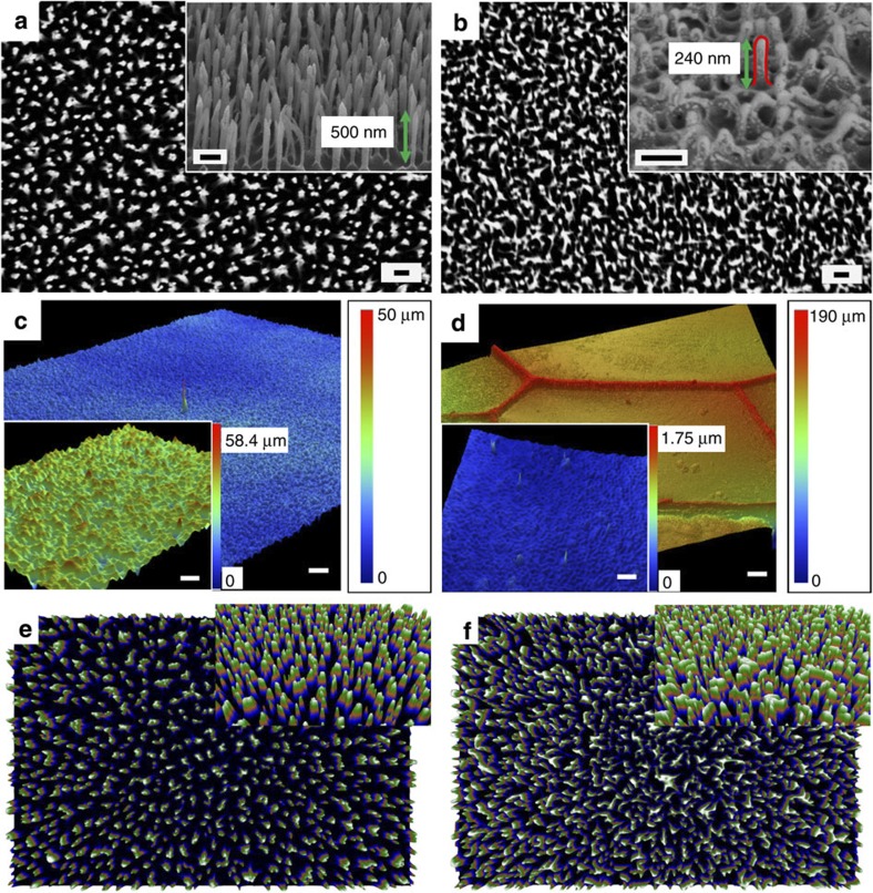
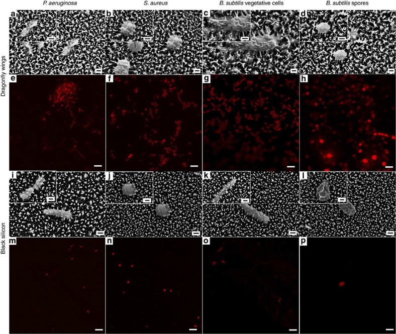
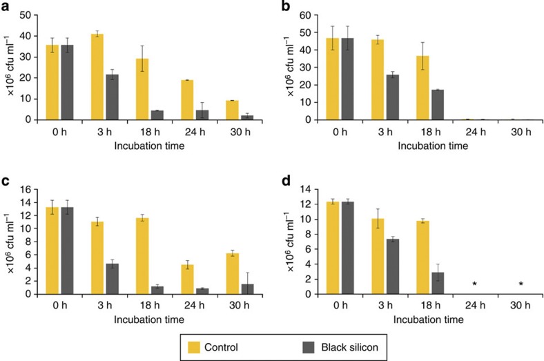
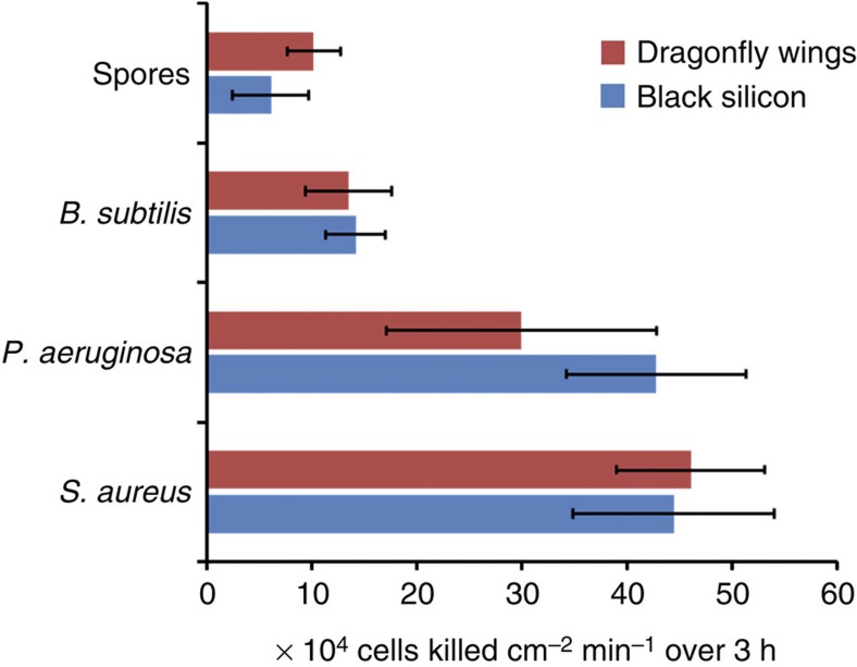
Black silicon is a synthetic nanomaterial that contains high aspect ratio nanoprotrusions on its surface, produced through a simple reactive-ion etching technique for use in photovoltaic applications. Surfaces with high aspect-ratio nanofeatures are also common in the natural world, for example, the wings of the dragonfly Diplacodes bipunctata. Here we show that the nanoprotrusions on the surfaces of both black silicon and D. bipunctata wings form hierarchical structures through the formation of clusters of adjacent nanoprotrusions. These structures generate a mechanical bactericidal effect, independent of chemical composition. Both surfaces are highly bactericidal against all tested Gram-negative and Gram-positive bacteria, and endospores, and exhibit estimated average killing rates of up to ~450,000 cells min(-1) cm(-2). This represents the first reported physical bactericidal activity of black silicon or indeed for any hydrophilic surface. This biomimetic analogue represents an excellent prospect for the development of a new generation of mechano-responsive, antibacterial nanomaterials.


Figures

Figure 1
Figure 1. Characterization of black silicon and D. bipunctata wings.
Scanning electron micrographs of the upper surface of (a) bSi and (b) dragonfly forewings at × 35,000 magnification demonstrate the surface patterns of the two samples. Scale bars, 200 nm. Micrographs tilted at an angle of 53° (inset) show sharper nanopillars of black silicon distinct from one another and approximately twice the height of those of the dragonfly wing. Optical profilometry shows the nanoprotrusions of (c) bSi and (d) dragonfly forewings. Scale bars, 50 μm; inset, 2 μm. Three-dimensional reconstructions based on a displacement map technique further highlight the differences and similarities of (e) bSi and (f) dragonfly forewings.
Figure 2
Figure 2. Cell morphology on dragonfly wings and black silicon.
SEM images of P. aeruginosa, S. aureus, B. subtilis vegetative cells and B. subtilis spores appear to be significantly disrupted through interaction with both the dragonfly wing (ad) and bSi (il). Scale bars, 200 nm. Confocal laser scanning micrographs confirm that disruption by dragonfly wing (eh) and bSi (mp) was lethal to the cells; non-viable bacterial cells and spores were stained with propidium iodide (red), whereas the living cells were stained with SYTO 9 (green). All cells appeared red, indicating the high efficiency of surfaces in inactivating the bacteria. Scale bars, 5 μm.
Figure 3
Figure 3. Bactericidal activity of black silicon surfaces.
Counts of viable cells of (a) P. aeruginosa, (b) S. aureus, (c) B. subtilis and (d) B. subtilis spores remaining in suspension when incubated with bSi and control suspensions for up to 30 h. At longer time intervals (24 and 30 h) cell numbers tended to also decrease in the controls due to the nutrient-poor medium. Data for spores in suspension after 24 and 30 h are not available due to the germination of spores after 18 h. Error bars are s.d. values.
Figure 4
Figure 4. Bactericidal efficiency of black silicon and dragonfly wings.
The bactericidal efficiency of both the bSi and D. bipunctata wing surfaces over the first 3 h of incubation was calculated as the number of cells killed per square centimetre of projected sample surface area, per minute of incubation time. The number of cells killed was determined by subtracting the number of surviving cells from the number of cells remaining in controls at the corresponding time interval. The uncertainty values represent the natural variability associated with biological systems, and were calculated based on standard deviations in colony forming units. Error bars are based on s.d. values.

References

    1. Chen F. et al. Anisotropic wetting on microstrips surface fabricated by femtosecond laser. Langmuir 27, 359–365 (2011). - PubMed
    1. Guo Z., Liu W. & Su B.-L. Superhydrophobic surfaces: from natural to biomimetic to functional. J. Colloid Interf. Sci. 353, 335–355 (2011). - PubMed
    1. Di Mundo R. et al. Cell adhesion on nanotextured slippery superhydrophobic substrates. Langmuir 27, 4914–4921 (2011). - PubMed
    1. Truong V. K. et al. Air-directed attachment of coccoid bacteria to the surface of superhydrophobic lotus-like titanium. Biofouling 28, 539–550 (2012). - PubMed
    1. Ivanova E. P. et al. Molecular organization of the nanoscale surface structures of the dragonfly Hemianax papuensis wing epicuticle. PLoS One 8, (2013). - PMC - PubMed

Publication types