Review
. 2014 Feb;34(2):185-99.
doi: 10.1038/jcbfm.2013.203. Epub 2013 Nov 27.

Hemorrhagic transformation after ischemic stroke in animals and humans

Affiliations
Review

Hemorrhagic transformation after ischemic stroke in animals and humans

Glen C Jickling et al. J Cereb Blood Flow Metab. 2014 Feb.

Abstract

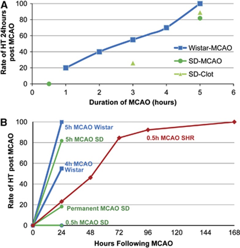
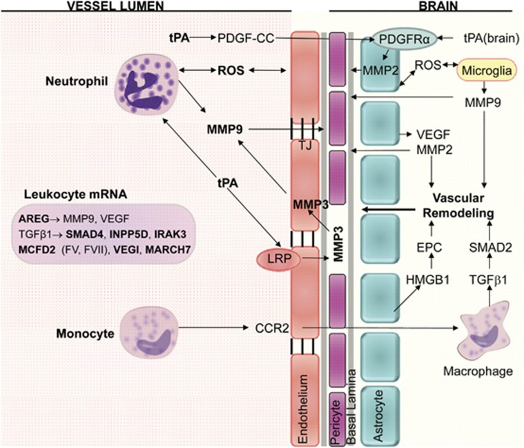
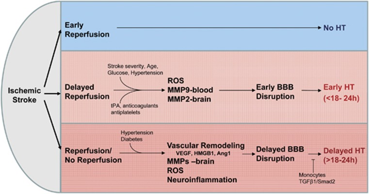
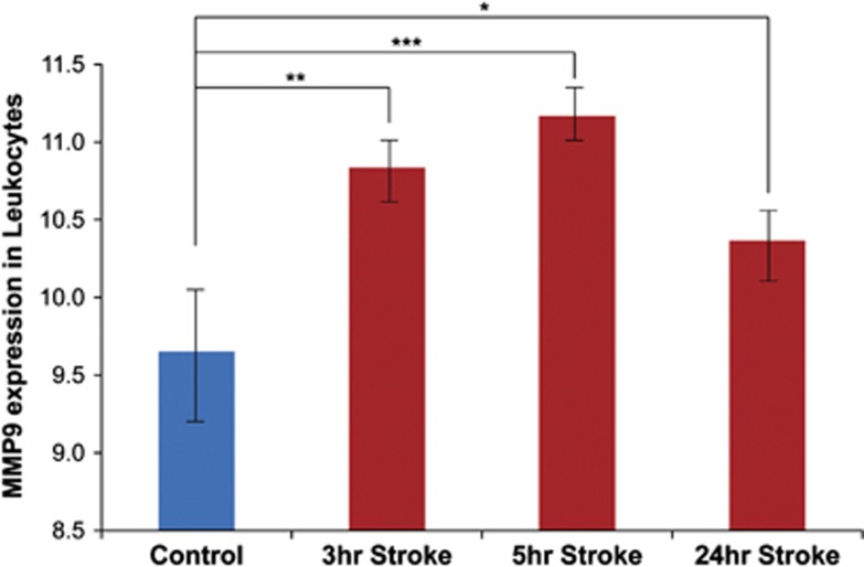
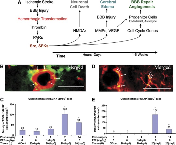
Hemorrhagic transformation (HT) is a common complication of ischemic stroke that is exacerbated by thrombolytic therapy. Methods to better prevent, predict, and treat HT are needed. In this review, we summarize studies of HT in both animals and humans. We propose that early HT (<18 to 24 hours after stroke onset) relates to leukocyte-derived matrix metalloproteinase-9 (MMP-9) and brain-derived MMP-2 that damage the neurovascular unit and promote blood-brain barrier (BBB) disruption. This contrasts to delayed HT (>18 to 24 hours after stroke) that relates to ischemia activation of brain proteases (MMP-2, MMP-3, MMP-9, and endogenous tissue plasminogen activator), neuroinflammation, and factors that promote vascular remodeling (vascular endothelial growth factor and high-moblity-group-box-1). Processes that mediate BBB repair and reduce HT risk are discussed, including transforming growth factor beta signaling in monocytes, Src kinase signaling, MMP inhibitors, and inhibitors of reactive oxygen species. Finally, clinical features associated with HT in patients with stroke are reviewed, including approaches to predict HT by clinical factors, brain imaging, and blood biomarkers. Though remarkable advances in our understanding of HT have been made, additional efforts are needed to translate these discoveries to the clinic and reduce the impact of HT on patients with ischemic stroke.


Figures

Figure 1
Figure 1
(A) The rate of hemorrhagic transformation (HT) increases with longer durations of cerebral ischemia followed by reperfusion. Middle cerebral artery occlusions (MCAOs) lasting 1 to 5.5 hours were induced in Wistar and Sprague Dawley (SD) rats. After reperfusion, the rate of HT was determined 24 hours after stroke onset., , , (B) The rate of HT as a function of MCAO duration. Increased duration of MCAO increases the rate of HT in several rat stroke models. Evaluation beyond 24 hours of stroke onset identifies additional HT in the spontaneously hypertensive rat (SHR). Note that a 0.5-hour MCAO in an SD rat does not produce HT at 24 hours, whereas a 0.5-hour MCAO in the SHR does produce HT with a 100% HT rate at 7 days after stroke onset.
Figure 2
Figure 2
Diagram of cell types and molecules associated with hemorrhagic transformation (HT) after ischemic stroke. Neutrophils are a major source of MMP-9 within the first 18 to 24 hours of stroke onset, and brain is a major source of MMP-9 at >18 to 24 hours after stroke onset. Exogenous tPA can act on neutrophils to increase MMP-9. Endogenous tPA can act on endothelial cell lipoprotein receptor protein (LRP) to increase MMP-3, and on PDGF-CC to increase MMP-2 via astrocyte PDGFRα receptors. Monocytes bind CCR2, enter brain where TGFβ1 induction of SMAD2 stabilizes the blood–brain barrier (BBB) and prevents HT for 1 to 7 days after stroke. AREG, amphiregulin; CCR2, C-C chemokine receptor type 2; EPC, endothelial progenitor cell; HMGB1, high-mobility-group-box-1; INPP5D, phosphatidylinositol-3,4,5-trisphosphate 5-phosphatase 1; IRAK3, interleukin-1 receptor-associated kinase 3; MARCH7, membrane-associated ring finger (C3HC4) 7; MCFD2, multiple coagulation factor D2; MMP, matrix metalloproteinase; PDGF-CC, platelet-derived growth factor CC; PDGFRα, platelet-derived growth factor receptor alpha; ROS, reactive oxygen species; SMAD, mothers against decapentaplegic homolog; TGFβ1, transforming growth factor β1; tPA, tissue plasminogen activator; VEGF, vascular endothelial growth factor; VEGI, vascular endothelial growth inhibitor.
Figure 3
Figure 3
Early reperfusion reduces the risk of hemorrhagic transformation (HT). Delayed reperfusion increases the BBB disruption and the risk of HT. ROS, leukocyte-derived MMP-9, and brain-derived MMP-2 have an important role in producing early BBB disruption and early HT. In contrast, brain-derived vascular remodeling, MMPs (MMP-9, MMP-2, and MMP-3), other brain proteases (plasmin, endogenous tPA, urokinase (uPA), and cathepsins), ROS, and neuroinflammation contribute to delayed BBB and delayed HT. A subset of monocytes that enter brain may prevent delayed HT. BBB, blood–brain barrier; MMP, matrix metalloproteinase; ROS, reactive oxygen species; SMAD, mothers against decapentaplegic homolog; TGFβ1, transforming growth factor β1; tPA, tissue plasminogen activator; VEGF, vascular endothelial growth factor; HMGB1, high-mobility-group-box-1.
Figure 4
Figure 4
Matrix metalloproteinase-9 (MMP-9) mRNA levels in blood of patients with ischemic stroke. MMP-9 mRNA was elevated at 3 and 5 hours after stroke compared with controls, with a return closer to baseline by 24 hours (*P=0.02; **P=9.9 × 10⁻⁵, ***P=8.6 × 10⁻⁸).
Figure 5
Figure 5
(A) Hemorrhagic transformation (HT) activates src and src family kinases (SFKs) via thrombin induction of protease-activated receptors (PARs). Within the first hours after stroke SFKs activate N-methyl D-aspartate receptors (NMDAr) and induce neuronal cell death. Within the first few days of hemorrhage, SFKs activate matrix metalloproteinases (MMPs), vascular endothelial growth factor (VEGF), and other molecules involved in blood–brain barrier (BBB) injury and cerebral edema. Beginning around 1 week after hemorrhage, SFKs activate cell-cycle genes in progenitor cells to form new brain endothelial cells and astrocytes that are involved in BBB repair and angiogenesis. (B) HT produces thrombin that induces the birth of new cerebral endothelial cells at day 7 and day 14 in rat hippocampus (green=BrdU (bromodeoxyuridine, a marker of cell proliferation); red=RECA-1 (rat endothelial cell antigen-1, endothelial cell marker); yellow=merged). (C) There is an increase in new cerebral endothelial cells (BrdU+ RECA-1+) in the hippocampus (LMol layer) on days 7 and 14 after thrombin injection. **P<0.01 (Cont=control; PP2=non-specific src family kinase inhibitor; Throm=thrombin; (D) HT produces thrombin that induces the birth of new perivascular astrocytes at day 7 and day 14 in rat hippocampus (green=BrdU; red=GFAP (green fluorescence protein, astrocyte marker); yellow=merged). (E) There is an increase in new perivascular astrocytes (BrdU+ GFAP+) in the hippocampus (LMol layer) on days 7 and 14 after thrombin injection. **P<0.01 (figures from Liu et al).

References

    1. Terruso V, D'Amelio M, Di Benedetto N, Lupo I, Saia V, Famoso G, et al. Frequency and determinants for hemorrhagic transformation of cerebral infarction. Neuroepidemiology. 2009;33:261–265. - PubMed
    1. Beslow LA, Smith SE, Vossough A, Licht DJ, Kasner SE, Favilla CG, et al. Hemorrhagic transformation of childhood arterial ischemic stroke. Stroke. 2011;42:941–946. - PMC - PubMed
    1. Fiorelli M, Bastianello S, von Kummer R, del Zoppo GJ, Larrue V, Lesaffre E, et al. Hemorrhagic transformation within 36 hours of a cerebral infarct: relationships with early clinical deterioration and 3-month outcome in the European Cooperative Acute Stroke Study I (ECASS I) cohort. Stroke. 1999;30:2280–2284. - PubMed
    1. Khatri P, Wechsler LR, Broderick JP. Intracranial hemorrhage associated with revascularization therapies. Stroke. 2007;38:431–440. - PubMed
    1. Berger C, Fiorelli M, Steiner T, Schabitz WR, Bozzao L, Bluhmki E, et al. Hemorrhagic transformation of ischemic brain tissue: asymptomatic or symptomatic. Stroke. 2001;32:1330–1335. - PubMed

Publication types

MeSH terms