. 2014 Jan;143(1):19-31.
doi: 10.1007/s10549-013-2779-4. Epub 2013 Nov 27.

Prostaglandin E receptor EP4 is a therapeutic target in breast cancer cells with stem-like properties

Affiliations

Prostaglandin E receptor EP4 is a therapeutic target in breast cancer cells with stem-like properties

Namita Kundu et al. Breast Cancer Res Treat. 2014 Jan.

Abstract

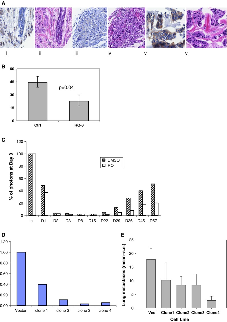
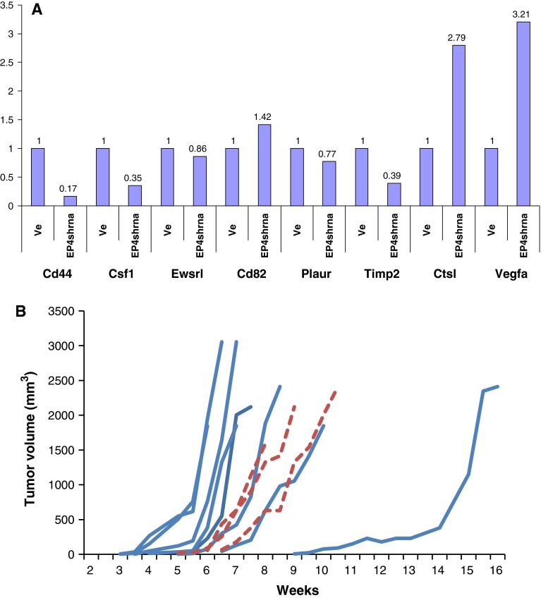
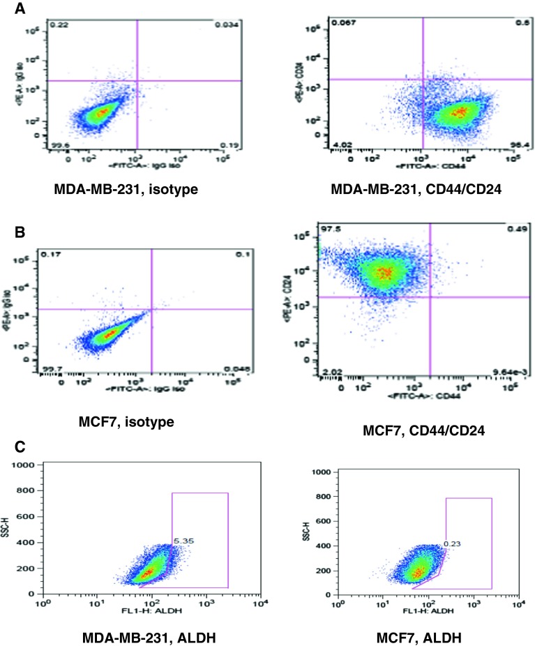
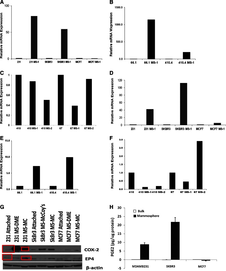
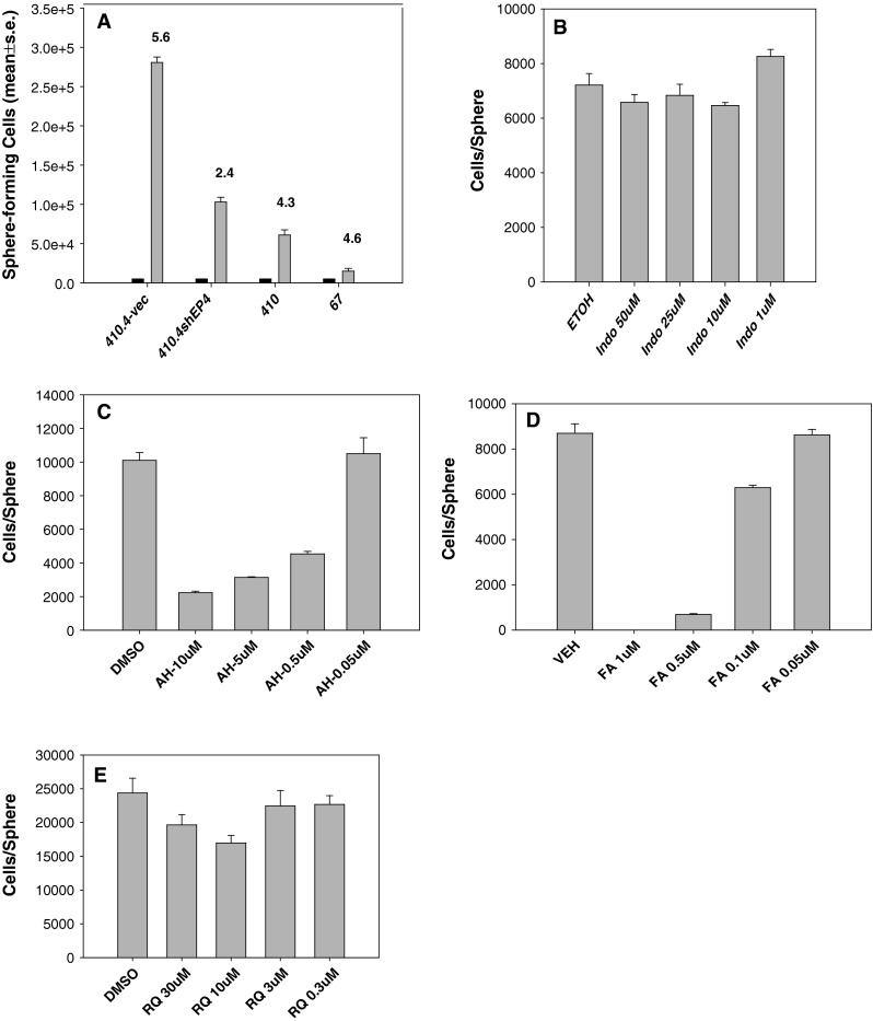
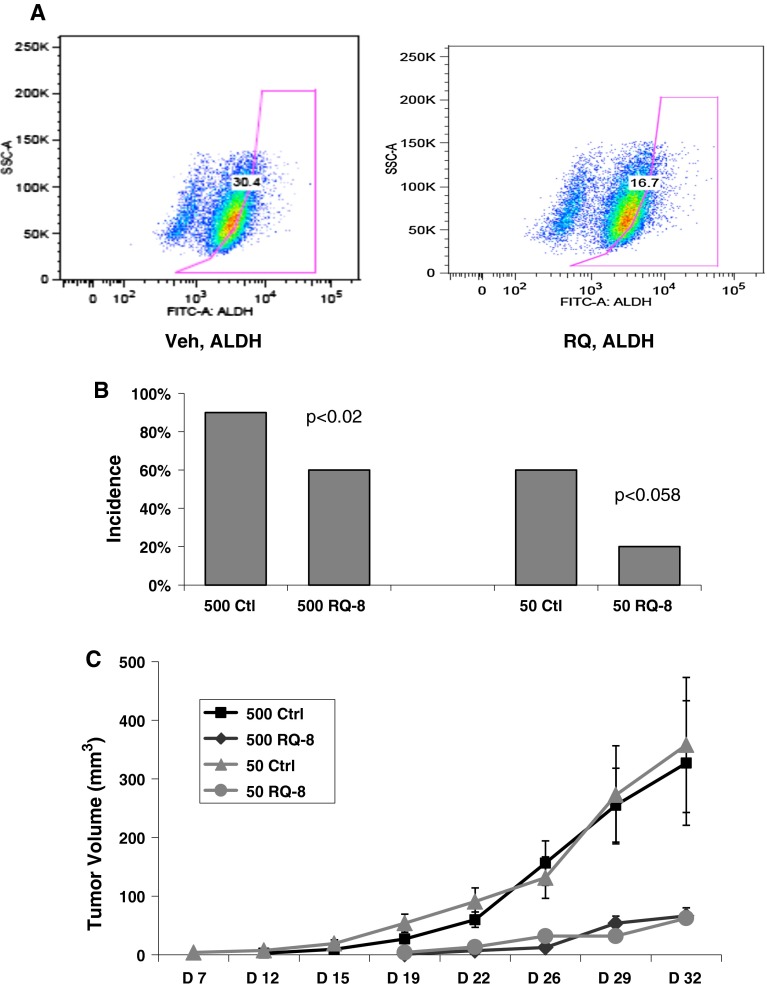
The cyclooxygenase pathway is strongly implicated in breast cancer progression but the role of this pathway in the biology of breast cancer stem/progenitor cells has not been defined. Recent attention has focused on targeting the cyclooxygenase 2 (COX-2) pathway downstream of the COX-2 enzyme by blocking the activities of individual prostaglandin E (EP) receptors. Prostaglandin E receptor 4 (EP4) is widely expressed in primary invasive ductal carcinomas of the breast and antagonizing this receptor with small molecule inhibitors or shRNA directed to EP4 inhibits metastatic potential in both syngeneic and xenograft models. Breast cancer stem/progenitor cells are defined as a subpopulation of cells that drive tumor growth, metastasis, treatment resistance, and relapse. Mammosphere-forming breast cancer cells of human (MDA-MB-231, SKBR3) or murine (66.1, 410.4) origin of basal-type, Her-2 phenotype and/or with heightened metastatic capacity upregulate expression of both EP4 and COX-2 and are more tumorigenic compared to the bulk population. In contrast, luminal-type or non-metastatic counterparts (MCF7, 410, 67) do not increase COX-2 and EP4 expression in mammosphere culture. Treatment of mammosphere-forming cells with EP4 inhibitors (RQ-15986, AH23848, Frondoside A) or EP4 gene silencing, but not with a COX inhibitor (Indomethacin) reduces both mammosphere-forming capacity and the expression of phenotypic markers (CD44(hi)/CD24(low), aldehyde dehydrogenase) of breast cancer stem cells. Finally, an orally delivered EP4 antagonist (RQ-08) reduces the tumor-initiating capacity and markedly inhibits both the size of tumors arising from transplantation of mammosphere-forming cells and phenotypic markers of stem cells in vivo. These studies support the continued investigation of EP4 as a potential therapeutic target and provide new insight regarding the role of EP4 in supporting a breast cancer stem cell/tumor-initiating phenotype.


Conflict of interest statement

The authors declare no competing interests.

Figures

Fig. 1
Fig. 1
a A tissue microarray was prepared containing 44 invasive ductal carcinoma of the breast. EP4 and H&E by immunohistochemistry. (i) Benign lobule, EP4, 1+; (ii) H&E; (iii) invasive ductal carcinoma, EP4, 1+; (iv) H&E; (v) invasive ductal carcinoma, EP4, 3+; (vi) H&E. b Line 410.4 tumor cells injected proximal to the mammary fat pad of Balb/cByJ female mice treated with vehicle or RQ-08 (30 mg/kg/day). When tumors measured 18 mm in diameter, mice were euthanized and surface lung tumor colonies enumerated. Mean ± SE, P = 0.04. c MDA-MB-231-luciferase cells treated with RQ-15986 (3.0 μM/l) or DMSO vehicle and injected i.v. into groups of five Balb/SCID mice and live animal imaging carried out at 5 min and at the days indicated. Data expressed as percent photons detected relative to day 0. d Line 66.1 cells transfected with plasmid expressing shEP4 or vector; stable clones were derived and EP4 expression characterized by qPCR. e Cell lines from d injected i.v. into 5–10 Balb/cByJ female mice and surface lung tumor colonies quantified. Mean ± SE, P < 0.01
Fig. 2
Fig. 2
a mRNA isolated from 66.1-vector and 66.1shEP4 cells and analyzed for expression of metastasis-related genes by metastasis pcr array. b 410.4 cells grown in standard culture or as mammospheres and on day 10 cells were harvested and 100 MS-1 or standard bulk culture cells injected into the mammary fat pad of Balb/cByJ female mice. Tumor growth monitored by caliper and expressed as tumor volume. Solid line = 100 MS-1 cells; dashed line = 100 bulk population cells
Fig. 3
Fig. 3
a, b MDA-MB-231 and MCF7 cells grown in standard culture conditions and analyzed for CD44 and CD24 expression by flow cytometry. Isotype control (left panel), double stain for CD44 and CD24 (right panel). c Aldefluor assay of MDA-MB-231 (left panel) and MCF7 (right panel) cells
Fig. 4
Fig. 4
a MDA-MB-231, SKBR3, or MCF7 cells or b 66.1 or 410.4 or c 410 or 67 cells grown as MS-1, MS-2, or bulk cultures. mRNA harvested and analyzed for human or murine EP4 by qPCR. df The same cells analyzed for COX-2 mRNA expression. g Cell lysates of MDA-MB-231, SKBR3, or MCF7 cells grown as standard culture or in mammosphere (MS) assay and maintained in either DME media or mammocult (MC) media and immunoblotted for COX-2, EP4, or β-actin. h MDA-MB-231, SKBR3, or MCF7 cells grown as attached (open bar) or mammosphere (closed bar) in DME; conditioned media analyzed for PGE2 by ELISA and expressed as Mean ± SE, pg/μg protein
Fig. 5
Fig. 5
a 5 × 103 MS-1 cells of 410.4-vec, 410.4shEP4, 410 or 67 cells (black bars) were re-plated in secondary (MS-2) cultures and on day 10, sphere number and cellularity were determined for MS-2 cultures. Mean ± SE. be MDA-MB-231 cells placed in mammosphere culture and on day 2, treated with Indomethacin, AH23848, Frondoside A or RQ-15986 at the indicated concentrations. On day 10, sphere number and cellularity were determined
Fig. 6
Fig. 6
a MDA-MB-231 cells in mammosphere culture were treated with vehicle (left panel) or RQ-15986 (10 μM/l, right panel) on day 8 and 2 days later, Aldefluor positive cells were determined. b Five hundred or 50 of 410.4 MS-1 cells were injected into groups of 10 Balb/cByJ female mice. On day +7, RQ-08 or vehicle administered by gavage (30 mg/kg/day) and tumor incidence on day +46 is plotted. c Tumor volume for the same mice as in b that developed palpable lesions. Mean ± SE

References

    1. Ristimaki A, Sivula A, Lundin J, Lundin M, Salminen T, Haglund C, et al. Prognostic significance of elevated cyclooxygenase-2 expression in breast cancer. Cancer Res. 2002;62:632–635. - PubMed
    1. Kerikowske K, Molinaro AM, Gauthier ML, Berman HK, Waldman F, Bennington J, Sanchez H, et al. Biomarker expression and risk of subsequent tumors after initial ductal carcinoma in situ diagnosis. J Natl Cancer Inst. 2010;102:627–637. doi: 10.1093/jnci/djq101. - DOI - PMC - PubMed
    1. Reader JC, Holt D, Fulton AM. Prostaglandin E2 EP receptors as therapeutic targets in breast cancer. Cancer Metastasis Rev. 2011;30:449–463. doi: 10.1007/s10555-011-9303-2. - DOI - PMC - PubMed
    1. Mutoh M, Watanabe K, Kitamura T, Shoji Y, Takahashi M, Kawamori T, et al. Involvement of prostaglandin E receptor subtype EP4 in colon carcinogenesis. Cancer Res. 2002;62:28–32. - PubMed
    1. Terada N, Shimizu Y, Kamba T, Inoue T, Maeno A, Kobayashi T, Nakamura E. Identification of EP4 as a potential target for the treatment of castration-resistant prostate cancer using a novel xenograft model. Cancer Res. 2010;70:1606–1615. doi: 10.1158/0008-5472.CAN-09-2984. - DOI - PubMed

Publication types

MeSH terms

Substances