Comparative Study
. 2014 Apr 15;23(8):2055-77.
doi: 10.1093/hmg/ddt600. Epub 2013 Nov 26.

Functional interaction of Parkinson's disease-associated LRRK2 with members of the dynamin GTPase superfamily

Affiliations
Comparative Study

Functional interaction of Parkinson's disease-associated LRRK2 with members of the dynamin GTPase superfamily

Klodjan Stafa et al. Hum Mol Genet. .

Abstract

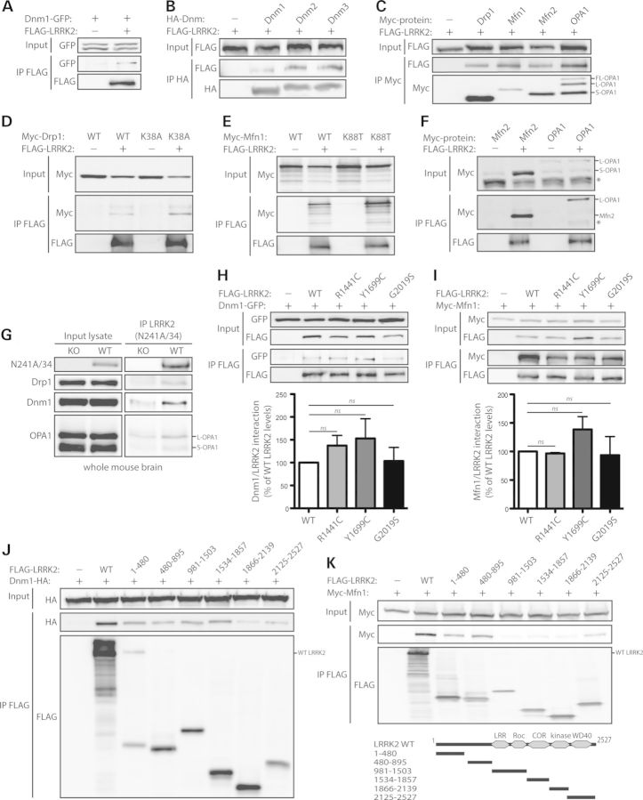
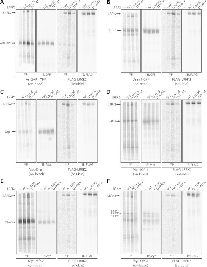
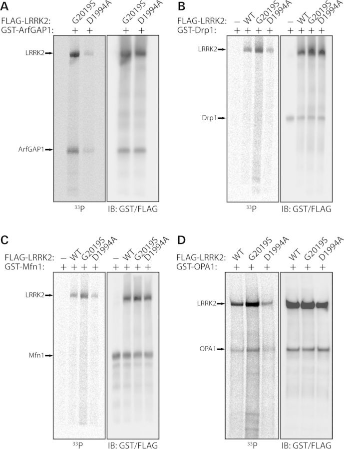
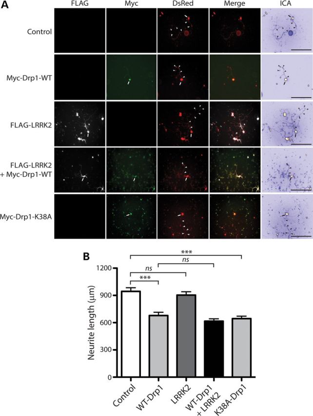
Mutations in LRRK2 cause autosomal dominant Parkinson's disease (PD). LRRK2 encodes a multi-domain protein containing GTPase and kinase domains, and putative protein-protein interaction domains. Familial PD mutations alter the GTPase and kinase activity of LRRK2 in vitro. LRRK2 is suggested to regulate a number of cellular pathways although the underlying mechanisms are poorly understood. To explore such mechanisms, it has proved informative to identify LRRK2-interacting proteins, some of which serve as LRRK2 kinase substrates. Here, we identify common interactions of LRRK2 with members of the dynamin GTPase superfamily. LRRK2 interacts with dynamin 1-3 that mediate membrane scission in clathrin-mediated endocytosis and with dynamin-related proteins that mediate mitochondrial fission (Drp1) and fusion (mitofusins and OPA1). LRRK2 partially co-localizes with endosomal dynamin-1 or with mitofusins and OPA1 at mitochondrial membranes. The subcellular distribution and oligomeric complexes of dynamin GTPases are not altered by modulating LRRK2 in mouse brain, whereas mature OPA1 levels are reduced in G2019S PD brains. LRRK2 enhances mitofusin-1 GTP binding, whereas dynamin-1 and OPA1 serve as modest substrates of LRRK2-mediated phosphorylation in vitro. While dynamin GTPase orthologs are not required for LRRK2-induced toxicity in yeast, LRRK2 functionally interacts with dynamin-1 and mitofusin-1 in cultured neurons. LRRK2 attenuates neurite shortening induced by dynamin-1 by reducing its levels, whereas LRRK2 rescues impaired neurite outgrowth induced by mitofusin-1 potentially by reversing excessive mitochondrial fusion. Our study elucidates novel functional interactions of LRRK2 with dynamin-superfamily GTPases that implicate LRRK2 in the regulation of membrane dynamics important for endocytosis and mitochondrial morphology.


Figures

Figure 1.
Figure 1.
LRRK2 commonly interacts with members of the dynamin GTPase superfamily. (A) FLAG-tagged human LRRK2 interacts with GFP-tagged human Dnm1 following immunoprecipitation (IP) with anti-FLAG antibody from HEK-293T cells. (B) HA-tagged mouse Dnm1, Dnm2 and Dnm3 interact with FLAG-LRRK2 following IP with anti-HA antibody. (C) Myc-tagged Drp1, Mfn1, Mfn2 and OPA1 interact with FLAG-LRRK2 following IP with anti-myc antibody. (D–F) FLAG-LRRK2 interacts with (D) Myc-tagged WT and K38A Drp1, (E) WT and K88T Mfn1 and (F) Mfn2 and OPA1, following IP with anti-FLAG antibody from HEK-293T cells. (G) LRRK2 interacts with Drp1, Dnm1 and OPA1 in whole brain extracts from WT mice but not LRRK2 knockout (KO) mice following IP with anti-LRRK2 antibody (clone N241A/34). Data are representative of two-independent experiments. (H) Dnm1-GFP and (I) Myc-Mfn1 interact with WT and PD-associated mutant forms of FLAG-LRRK2 following IP with anti-FLAG antibody from HEK-293T cells. Densitometric analysis reveals no significant differences in the interactions of Dnm1 or Mfn1 with R1441C, Y1699C and G2019S LRRK2 compared with WT LRRK2. Data represent the level of interaction of Dnm1 or Mfn1 with LRRK2 expressed as a percent of the interaction with WT LRRK2. The levels of Dnm1 or Mfn1 IP were first normalized to their respective input levels, and then further normalized to LRRK2 IP levels. Bars represent the mean ± SEM (n = 3 experiments). n.s., non-significant. (J and K) Domain mapping reveals the interaction of HA-Dnm1 with full-length (WT) LRRK2, residues 1–480, 480–895, 981–1503 and 1534–1857 of LRRK2, and to a lesser extent other LRRK2 domains (J), whereas Myc-Mfn1 interacts with full-length LRRK2 and residues 1–480, 480–895 and 2125–2527 of LRRK2 (K) following IP with anti-FLAG antibody. (K) Domain organization of LRRK2 deletion mutants is indicated. Data are representative of at least three-independent experiments.
Figure 2.
Figure 2.
LRRK2 partially co-localizes with Dnm1 at early endosomes in neural cells. (A–C) Confocal fluorescence microscopy reveals the partial co-localization of FLAG-tagged human LRRK2 (WT, G2019S and R1441C) with HA-tagged human Dnm1 at RFP-Rab5-positive early endosomal vesicles in human SH-SY5Y neural cells. Cytofluorograms and co-localization coefficients (Rcoloc; mean ± SEM, n = 10–12 cells/condition) reveal the extent of co-localization between LRRK2 and Dnm1 fluorescence signals. (A–D) Confocal images, cytofluorograms and co-localization coefficients revealing the effect of overexpressing human LRRK2 variants on the degree of co-localization of Dnm1 and Rab5, relative to control cells transfected with empty vector (pcDNA3.1). Confocal images are taken from a single z-plane at 0.1 µm thickness. Images are representative of multiple cells from at least two-independent transfection experiments. Scale bars: 10 µm. (E and F) Graphs indicating co-localization coefficients (mean ± SEM, n = 10–12 cells) for (E) Dnm1 and LRRK2 variants and (F) Dnm1 and Rab5. The G2019S mutation significantly increases the co-localization of LRRK2 with Dnm1 (E), and reduces the co-localization of Dnm1 with Rab5-positive endosomes (F). *P < 0.05 and **P < 0.01 by one-way ANOVA with Newman–Keuls post hoc analysis, as indicated. n.s., non-significant.
Figure 3.
Figure 3.
LRRK2 partially co-localizes with dynamin-related GTPases at mitochondria in cortical neurons. Confocal fluorescence microscopy reveals the partial co-localization of FLAG-tagged human LRRK2 with Myc-tagged Drp1 (WT and K38A), Mfn1 (WT and K88T), Mfn2 and OPA1 at mito-RFP-positive mitochondria in rat primary cortical neurons. Cytofluorograms and co-localization coefficients (Rcoloc; mean ± SEM, n ≥ 5 cells) reveal the extent of co-localization of LRRK2 with Drp1, Mfn1, Mfn2 or OPA1 fluorescence signals. The degree of co-localization of Drp1, Mfn1, Mfn2 and OPA1 with mito-RFP fluorescence signals is also indicated for comparison. Confocal images are taken from a single z-plane at 0.1 µm thickness. Images are representative of multiple neurons from at least two-independent transfection experiments. Scale bars: 10 µm.
Figure 4.
Figure 4.
Subcellular distribution and native complexes of dynamin GTPases are not altered by LRRK2 in mouse brain. (A) Subcellular fractionation of cerebral cortex tissue derived from human G2019S LRRK2 transgenic and non-transgenic mice. Dnm1 and Drp1 are broadly distributed across multiple membrane and soluble fractions, whereas Mfn2 and OPA1 are enriched in heavy membrane (P2) and synaptosomal membrane (LP1) fractions. LRRK2 is broadly detected with enrichment in light membrane/microsomal (P3), synaptosomal LP1 and synaptic vesicle-enriched (LP2) membrane fractions. The distribution of the synaptic vesicle-associated protein, synaptophysin 1, demonstrates enrichment of membranes in P2, P3, LP1 and LP2 fractions, whereas Mfn2 and OPA1 indicate enrichment of mitochondria in P2 and LP1 fractions. (B) Native-PAGE and (C) SDS–PAGE analysis of equivalent cerebral cortex extracts derived from human G2019S LRRK2 transgenic (Tg) and non-transgenic (NTg) mice, and LRRK2 KO and WT mice, revealing similar oligomeric protein complexes for Dnm1, Drp1 and OPA1. (C) LRRK2 antibodies confirm the absence of LRRK2 in KO mice (mouse-selective N241A/34 antibody) and human G2019S LRRK2 expression in transgenic mice (human-selective MJFF4/c81-8 antibody; lower band = LRRK2; asterisk indicates non-specific upper band). (D and E) Size-exclusion chromatography on soluble whole brain extracts from WT and LRRK2 KO mice. Sequential fractions (#1–16, 0.5 ml) and total homogenates (WT or KO) were analyzed by western blotting with antibodies to Dnm1, Drp1 and LRRK2 (N241A/34), or β-tubulin as a loading control. The elution profile of Dnm1 and Drp1 is similar in WT and KO brains, whereas the elution profile of individual standards is indicated. LRRK2 antibody (N241A/34) confirms the absence of LRRK2 in KO mice. Blots are representative of duplicate experiments. Molecular mass markers are indicated in kDa.
Figure 5.
Figure 5.
Reduced levels of mature S-OPA1 in G2019S mutant PD brains. Western blot analysis of frontal cortex soluble fractions from human control, idiopathic PD (iPD) or G2019S LRRK2 PD subjects with antibodies to Dnm1, Drp1, OPA1 and Mfn2, or β-tubulin as a protein loading control. Densitometric analysis of Dnm1, Drp1, OPA1 (L-OPA1 and S-OPA1) and Mfn2 in idiopathic or G2019S PD brains compared with control brains are indicated. The levels of each protein were normalized to β-tubulin levels, and expressed as a percent of control subjects (mean ± SEM, n = 4 subjects/group). *P < 0.05 by one-way ANOVA with Newman–Keuls post hoc analysis, as indicated.
Figure 6.
Figure 6.
LRRK2 enhances the levels of GTP-bound Mfn1. Dynamin GTPases (Dnm1, Drp1, Mfn1, Mfn2 and OPA1) fail to significantly influence the steady-state levels of FLAG-tagged WT LRRK2 bound to GTP following pull-down assays with GTP-agarose from HEK-293T cell extracts. The specificity of LRRK2 GTP binding is indicated by the absence of binding of the GDP/GTP-binding-deficient LRRK2 mutant, T1348N (TN). Densitometric analysis reveals significantly enhanced steady-state levels of Mfn1 but not Dnm1 bound to GTP in the presence of LRRK2. Note that only Mfn1 and Dnm1 reveal detectable GTP binding when expressed alone compared with Drp1, Mfn2 and OPA1. Molecular mass markers are indicated in kDa. Data represent the level of LRRK2 (left) or Dnm1 and Mfn1 (right) GTP binding expressed as a percent of the levels of each protein alone. GTP-bound protein levels were normalized to input protein levels. Bars represent the mean ± SEM (n = 3 experiments). *P < 0.05 or **P < 0.002 by one-way ANOVA with Newman–Keuls post hoc analysis, as indicated. n.s., non-significant.
Figure 7.
Figure 7.
Dynamin GTPases are modestly phosphorylated by LRRK2 in vitro. In vitro kinase assays with [33P]-γ-ATP, soluble recombinant full-length FLAG-tagged LRRK2 variants (WT, G2019S or D1994A) and immunopurified ‘on-bead’ YFP-tagged ArfGAP1 (A), GFP-tagged Dnm1 (B), and Myc-tagged Drp1 (C), Mfn1 (D), Mfn2 (E) or OPA1 (F), derived by IP from transfected HEK-293T cells. Following kinase reactions, soluble LRRK2 and ‘on-bead’ substrates were separated and resolved on independent SDS–PAGE gels, as indicated. Western blot analysis with anti-GFP, anti-myc or anti-FLAG antibodies indicate equal loading of ArfGAP1, Dnm1, Drp1, Mfn1, Mfn2, OPA1 and LRRK2 proteins in each condition. Autoradiographs (33P) reveal the LRRK2-dependent phosphorylation of ArfGAP1, Dnm1, Drp1, Mfn1 and OPA1, with enhanced phosphorylation by G2019S LRRK2 compared with WT or kinase-inactive D1994A LRRK2. A soluble eluate from FLAG IPs (derived from non-transfected cells) was used as a control in each assay to assess background 33P incorporation for each substrate. LRRK2 autophosphorylation is also detected in these assays. Blots are representative of at least three-independent kinase experiments.
Figure 8.
Figure 8.
Recombinant OPA1 is phosphorylated by LRRK2 in vitro. In vitro kinase assays with [33P]-γ-ATP, recombinant soluble full-length FLAG-tagged LRRK2 variants (WT, G2019S or D1994A) and recombinant full-length GST-tagged ArfGAP1 (A), Drp1 (B), Mfn1 (C) or OPA1 (D). Western blot analyses with anti-GST and anti-FLAG antibodies indicate equal loading and positions of ArfGAP1, Drp1, Mfn1, OPA1 and LRRK2 proteins in each condition. Autoradiographs (33P) reveal the LRRK2-dependent phosphorylation of ArfGAP1 and OPA1, with enhanced phosphorylation by G2019S LRRK2 compared with WT or D1994A LRRK2. A soluble eluate from FLAG IPs (derived from non-transfected cells) was used as a control in each assay to assess background 33P incorporation for each substrate. LRRK2 autophosphorylation is also detected in these assays. Blots are representative of at least three-independent kinase experiments.
Figure 9.
Figure 9.
Mitochondrial dynamin GTPases are not required for LRRK2-induced toxicity in yeast. Yeast cells (BY4741 MATa), either WT or deletion mutants, were transformed with (A) galactose-inducible or (B) constitutively expressing (p426GPD) high-copy expression constructs containing human LRRK2 (residues 1300–2527). A corresponding empty vector (p426GAL1 or p426GPD) was used as a control. Cells were spotted onto SC-URA media containing glucose, galactose, glycerol or ethanol as the sole carbon source, as indicated, and incubated at 30°C for up to 6 days. Shown are 5-fold serial dilutions (from left to right) starting with equal numbers of cells. Data are representative of two independently transformed clones for each plasmid.
Figure 10.
Figure 10.
LRRK2 attenuates neurite shortening induced by Dnm1. (A) Primary cortical neurons were co-transfected with FLAG-tagged WT LRRK2, GFP-tagged Dnm1 and DsRed-Max constructs at a molar ratio of 10:10:1 at DIV 3 and fixed at DIV 7. Fluorescent microscopic images indicate the co-labeling of cortical neurons with combinations of FLAG-LRRK2, Dnm1-GFP and DsRed. DsRed images were pseudo-colored with ICA for neurite length measurements. Neuronal soma (arrows) and axonal processes (arrowheads) are indicated. Scale bars: 400 µm. (B) Analysis of DsRed-positive neurites reveals a marked shortening of axonal processes by Dnm1 expression alone, with a modest effect of WT LRRK2 expression alone, compared with control neurites (DsRed alone). Co-expression of WT LRRK2 and Dnm1 markedly attenuates the Dnm1-induced shortening of axonal processes. Bars represent axonal process length (mean ± SEM) expressed as a percent of DsRed alone (control) from ≥90 DsRed-positive neurons taken from at least three-independent experiments/cultures. *P < 0.05, **P < 0.01 or ***P < 0.001 by one-way ANOVA with Newman–Keuls post hoc analysis. (C) Western blot analysis with anti-FLAG, anti-GFP and anti-β-tubulin antibodies of cell extracts derived from rat primary cortical neurons at DIV 7 transiently expressing FLAG-LRRK2 and Dnm1-GFP. Densitometric analysis reveals a strong trend (P = 0.059 by unpaired Student's t-test) towards reduced Dnm1 levels in the presence of LRRK2. Graphs indicate LRRK2 (left) or Dnm1 (right) steady-state levels normalized to β-tubulin levels, expressed as a percent of each protein alone (mean ± SEM, n = 3 experiments). n.s., non-significant.
Figure 11.
Figure 11.
LRRK2 does not influence neurite shortening induced by Drp1. (A) Primary cortical neurons were co-transfected with FLAG-tagged WT LRRK2, Myc-tagged Drp1 (WT or K38A) and DsRed-Max constructs at a molar ratio of 10:10:1 at DIV 3 and fixed at DIV 7. Fluorescent microscopic images indicate the co-labeling of cortical neurons with combinations of FLAG-LRRK2, Myc-Drp1 and DsRed. DsRed images were pseudo-colored with ICA for neurite length measurements. Neuronal soma (arrows) and axonal processes (arrowheads) are indicated. Scale bars: 400 µm. (B) Analysis of DsRed-positive neurites reveals a marked shortening of axonal processes by WT or K38A Drp1 expression alone, with a negligible effect of WT LRRK2 expression alone, compared with control neurites (DsRed alone). Co-expression of WT LRRK2 and WT Drp1 fails to alter Drp1-induced shortening of axonal processes. Bars represent axonal process length (mean ± SEM) expressed as a percent of DsRed alone (control) from ≥90 DsRed-positive neurons taken from at least three-independent experiments/cultures. ***P < 0.001 by one-way ANOVA with Newman–Keuls post hoc analysis. n.s., non-significant.
Figure 12.
Figure 12.
LRRK2 rescues impaired neurite outgrowth induced by Mfn1. (A) Primary cortical neurons were co-transfected with FLAG-tagged LRRK2 (WT or G2019S), Myc-tagged Mfn1 and DsRed-Max constructs at a molar ratio of 10:10:1 at DIV 3 and fixed at DIV 7. Fluorescent microscopic images indicate the co-labeling of cortical neurons with combinations of FLAG-LRRK2, Myc-Mfn1 and DsRed. DsRed images were pseudo-colored with ICA for neurite length measurements. Neuronal soma (arrows) and axonal processes (arrowheads) are indicated. Scale bars: 400 µm. (B) Analysis of DsRed-positive neurites reveals a marked shortening of axonal processes by Mfn1 or G2019S LRRK2 expression alone, with a negligible effect of WT LRRK2 expression alone, compared with control neurites (DsRed alone). Co-expression of G2019S LRRK2 and Mfn1 rescues the Mfn1-induced shortening of axonal processes, whereas WT LRRK2 partially rescues the effects of Mfn1. Bars represent axonal process length (mean ± SEM) expressed as a percent of DsRed alone (control) from ≥90 DsRed-positive neurons taken from at least three-independent experiments/cultures. **P < 0.01 or ***P < 0.001 by one-way ANOVA with Newman–Keuls post hoc analysis. n.s., non-significant. (C) Western blot analysis with anti-FLAG, anti-myc and anti-β-tubulin antibodies of cell extracts derived from rat primary cortical neurons at DIV 7 transiently expressing FLAG-LRRK2 and Myc-Mfn1. The levels of LRRK2 or Mfn1 are not altered when expressed alone or together. Blots are representative of three-independent experiments.

References

    1. Biskup S., West A.B. Zeroing in on LRRK2-linked pathogenic mechanisms in Parkinson's disease. Biochim. Biophys. Acta. 2009;1792:625–633. - PMC - PubMed
    1. Healy D.G., Falchi M., O'Sullivan S.S., Bonifati V., Durr A., Bressman S., Brice A., Aasly J., Zabetian C.P., Goldwurm S., et al. Phenotype, genotype, and worldwide genetic penetrance of LRRK2-associated Parkinson's disease: a case-control study. Lancet Neurol. 2008;7:583–590. - PMC - PubMed
    1. Simon-Sanchez J., Schulte C., Bras J.M., Sharma M., Gibbs J.R., Berg D., Paisan-Ruiz C., Lichtner P., Scholz S.W., Hernandez D.G., et al. Genome-wide association study reveals genetic risk underlying Parkinson's disease. Nat. Genet. 2009;41:1308–1312. - PMC - PubMed
    1. Tsika E., Moore D.J. Mechanisms of LRRK2-mediated neurodegeneration. Curr. Neurol. Neurosci. Rep. 2012;12:251–260. - PubMed
    1. Cookson M.R. The role of leucine-rich repeat kinase 2 (LRRK2) in Parkinson's disease. Nat. Rev. Neurosci. 2010;11:791–797. - PMC - PubMed

Publication types

MeSH terms