. 2013 Dec 10;110(50):E4922-30.
doi: 10.1073/pnas.1314197110. Epub 2013 Nov 26.

Intestinal HIF2α promotes tissue-iron accumulation in disorders of iron overload with anemia

Affiliations

Intestinal HIF2α promotes tissue-iron accumulation in disorders of iron overload with anemia

Erik R Anderson et al. Proc Natl Acad Sci U S A. .

Abstract

Several distinct congenital disorders can lead to tissue-iron overload with anemia. Repeated blood transfusions are one of the major causes of iron overload in several of these disorders, including β-thalassemia major, which is characterized by a defective β-globin gene. In this state, hyperabsorption of iron is also observed and can significantly contribute to iron overload. In β-thalassemia intermedia, which does not require blood transfusion for survival, hyperabsorption of iron is the leading cause of iron overload. The mechanism of increased iron absorption in β-thalassemia is unclear. We definitively demonstrate, using genetic mouse models, that intestinal hypoxia-inducible factor-2α (HIF2α) and divalent metal transporter-1 (DMT1) are activated early in the pathogenesis of β-thalassemia and are essential for excess iron accumulation in mouse models of β-thalassemia. Moreover, thalassemic mice with established iron overload had significant improvement in tissue-iron levels and anemia following disruption of intestinal HIF2α. In addition to repeated blood transfusions and increased iron absorption, chronic hemolysis is the major cause of tissue-iron accumulation in anemic iron-overload disorders caused by hemolytic anemia. Mechanistic studies in a hemolytic anemia mouse model demonstrated that loss of intestinal HIF2α/DMT1 signaling led to decreased tissue-iron accumulation in the liver without worsening the anemia. These data demonstrate that dysregulation of intestinal hypoxia and HIF2α signaling is critical for progressive iron overload in β-thalassemia and may be a novel therapeutic target in several anemic iron-overload disorders.

Keywords: Epas1; HIF; Slc11a2; thalassemia.


Conflict of interest statement

Conflict of interest statement: S. Rivella is a consultant for Exigo Management Consultant, Isis, Bayer AG, BioMarin, Merganser Biotech, and Novartis Pharmaceuticals. He also holds an equity/ownership interest in Merganser Biotech, Inc. In addition, he is a coinventor for the patents US8058061 B2 C12N 20111115 and US7541179 B2 C12N 20090602. The consulting work and intellectual property of S. Rivella did not affect in any way the design, conduct, or reporting of this research.

Figures
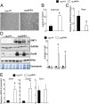
Fig. 1.
Fig. 1.
Apical iron absorption genes are increased early in a mouse model of β-thalassemia. (A) Enhanced Perls’ Prussian blue staining, (B) liver iron quantitation, and (C) qPCR analysis measuring liver hepcidin (Hepc) expression in 3-wk-old Hbbth3/+ and Hbb+/+ mice. Expression was normalized to β-actin. (D) Western blot analysis for DMT1, FPN1, and DcytB in 3-wk-old Hbbth3/+ and Hbb+/+ mice. DMT1 loading was normalized to glyceraldehyde-3-phosphate dehydrogenase (GAPDH) whereas FPN1 and DcytB were normalized to coomassie staining, and the blots were quantitated and presented as fold changes compared with Hbb+/+ mice (Right). (E) qPCR analysis measuring intestinal Dmt1, Fpn1, and Dcytb gene expression. Expression was normalized to β-actin. Four to eight mice were assessed per each group, and each bar graph represents the mean value ± SD. *P < 0.01.
Fig. 2.
Fig. 2.
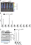
Mice with β-thalassemia have systemic and duodenal hypoxia. ODD-luc mice were crossed with Hbbth3/+ mice and examined at 3 wk of age. (A) In vivo analysis of hypoxia in Hbbth3/+ mice compared with littermate controls. (B) Tissue luciferase assays from duodenum (Duo), colon (Col), Liver (Liv), kidney (Kid), heart (Ht), spleen (Spl), pancreas (Pan), white adipose tissue (WAT), brown adipose tissue (BAT), and lung (Lu). Luciferase values [relative luciferase units (RLU)] were normalized to total protein. (C) Western blot for duodenal HIF2α, DMT1, and FPN1 expression. HIF2α expression was normalized to lamin B1, DMT1 expression was normalized to GAPDH, and FPN1 expression was normalized to coomassie staining, and the blots were quantitated and presented as fold changes compared with Hbb+/+ mice (Right). Three to five mice were assessed per each group, and each bar graph represents the mean value ± SD. *P < 0.01.
Fig. 3.
Fig. 3.
Disruption of intestinal HIF2α limits tissue-iron accumulation in a BMT-induced model of β-thalassemia. (A) Hif2αF/F and Hif2αΔIE mice were transplanted with Hbb+/+ or Hbbth3/+ bone marrow and analyzed 1 mo later for (B) intestinal Dmt1 and Dcytb mRNA expression and (C) liver iron content. mRNA expression was normalized to β-actin, and tissue-iron content was calculated per mg wet weight. Six to eight mice were assessed per each group, and each bar graph represents the mean value ± SD. *P < 0.01.
Fig. 4.
Fig. 4.
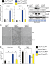
β-thalassemic mice crossed to Hif2αΔIE mice demonstrate decreased tissue-iron accumulation. (A) qPCR analysis measuring intestinal Dmt1 and Dcytb gene expression in 2-mo-old Hif2αF/F/Hbb+/+, Hif2αΔIE/Hbb+/+, Hif2αF/F/Hbbth3/+, and Hif2αΔIE/Hbbth3/+ mice. Expression was normalized to β-actin. (B) Western blot analysis measuring intestinal DMT1, DcytB, and FPN1 protein expression in 2-mo-old Hif2αF/F/Hbb+/+, Hif2αΔIE/Hbb+/+, Hif2αF/F/Hbbth3/+, and Hif2αΔIE/Hbbth3/+ mice. DMT1 loading was normalized to GAPDH whereas FPN1 and DcytB were normalized to coomassie staining The blots were then quantitated and presented as fold changes compared with Hbb+/+/Hif2αF/F mice (values below the blots). (C) Enhanced Perls’ Prussian blue staining, (D) iron quantitation from the livers, and (E) RBC values in 2-mo-old Hif2αF/F/Hbb+/+, Hif2αΔIE/Hbb+/+, Hif2αF/F/Hbbth3/+, and Hif2αΔIE/Hbbth3/+ mice. Six to eight mice were assessed per each group, and each bar graph represents the mean value ± SD. *P < 0.01. For Western quantitation, the mean values ± SD are represented. *P < 0.01 compared with Hif2αF/F/Hbb+/+ mice.
Fig. 5.
Fig. 5.
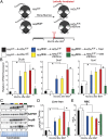
Disruption of HIF2α decreases tissue-iron accumulation in thalassemic mice with established iron overload. (A) Hif2αF/F, Hif2αΔIE, and Hif2αF/F;VilERcre mice were lethally irradiated and transplanted with Hbbth3/+ or Hbb+/+ bone marrow. Two months following BMT, mice were treated with vehicle or 1.5 mg of tamoxifen for 3 consecutive days and killed 2 mo later. (B) qPCR analysis measuring Dmt1, Dcytb, and Fpn1. Expression was normalized to β-actin. (C) Western blot analysis measuring intestinal DMT1, DcytB, and FPN1 protein expression. DMT1 loading was normalized to GAPDH whereas FPN1 and DcytB were normalized to Coomassie staining. The blots were quantitated and presented as fold changes compared with Hbb+/+ bone marrow transplanted into Hif2αF/F mice (values below the blots). (D) Iron quantitation from the liver and (E) RBC values. Six to eight mice were assessed per each group, and each bar graph represents the mean value ± SD. *P < 0.01. For Western quantitation, the mean values are represented. *P < 0.01 compared with Hbbth3/+ bone marrow transplanted into Hif2αF/F mice at 4 mo following BMT. TP < 0.01 compared with mice at 2 mo following BMT.
Fig. 6.
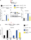
Fig. 6.
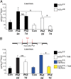
Intestinal disruption of HIF2α decreases hemolysis-induced liver iron accumulation. (A) Hif2αF/F and Hif2αΔIE mice were treated with vehicle (Con) or phenylhydrazine (PhZ), and liver iron was quantitated 2 and 7 d following treatment. (B) Hif2αF/F and Hif2αF/F;VilERcre mice were treated with vehicle (Con) or phenylhydrazine (PhZ), and liver iron was quantitated or the mice were additionally treated with 1.5 mg of tamoxifen for 3 consecutive days, and liver iron was quantitated 7 d following PhZ treatment. Four to eight mice were assessed per each group, and each bar graph represents the mean value ± SD. NS, not significant; *P < 0.01; #P < 0.05.
Fig. 7.
Fig. 7.
DMT1 is a critical downstream effector of HIF2α in anemic iron overload. (A) Dmt1F/+ and Dmt1F/+;VilERcre mice were treated with 1.5 mg of tamoxifen for 3 consecutive days and then were lethally irradiated and transplanted with Hbbth3/+ or Hbb+/+ bone marrow and analyzed 1 mo later. (B) qPCR analysis measuring Dmt1 with expression normalized to β-actin. (C) Western blot analysis measuring intestinal DMT1 with loading normalized to GAPDH. The blot was then quantitated and presented as fold change compared with Hbb+/+ bone marrow transplanted into Dmt1F/+ mice (values below the blots). (D) Iron quantitation from the liver of BMT mice. (E) Dmt1F/+ and Dmt1F/+;VilERcre mice were treated with vehicle (Con) or phenylhydrazine (PhZ), and liver iron was quantitated or the mice were additionally treated with 1.5 mg of tamoxifen for 3 consecutive days, and liver iron was quantitated 7 d following PhZ treatment. Six to eight mice were assessed per each group, and each bar graph represents the mean value ± SD. *P < 0.01, #P < 0.05. For Western quantitation, the mean values are represented. *P < 0.01 compared with Hbbth3/+ bone marrow transplanted into Dmt1F/+ mice.

References

    1. Gattermann N. The treatment of secondary hemochromatosis. Dtsch Arztebl Int. 2009;106(30):499–504, I. - PMC - PubMed
    1. Musallam KM, et al. Elevated liver iron concentration is a marker of increased morbidity in patients with β thalassemia intermedia. Haematologica. 2011;96(11):1605–1612. - PMC - PubMed
    1. Steinberg MHFB, Higgs DR, Nagel RL. Disorders of Hemoglobin: Genetics, Pathophysiology and Clincal Management. Cambridge, UK: Cambridge Univ Press; 2001.
    1. Heinrich HC, et al. Absorption of inorganic and food iron in children with heterozygous and homozygous beta-thalassemia. Z Kinderheilkd. 1973;115(1):1–22. - PubMed
    1. Pitcher CS, Williams HS, Parsonson A, Williams R. The measurement of iron absorption by the double isotope technique. Br J Haematol. 1965;11(6):633–641. - PubMed

Publication types

LinkOut - more resources