

Tissue kidney injury molecule-1 expression in the prediction of renal function for several years after kidney biopsy
- PMID: 24282337
- PMCID: PMC3824354
- DOI: 10.1155/2013/183246
Tissue kidney injury molecule-1 expression in the prediction of renal function for several years after kidney biopsy
Abstract
Objectives: Retrospective study was designed to examine the importance of tissue kidney injury molecule-1 (KIM-1) expression in predicting kidney function in sixty patients (27 males) aged 34.15 ± 12.23 years with different kidney diseases over three years after kidney biopsy.
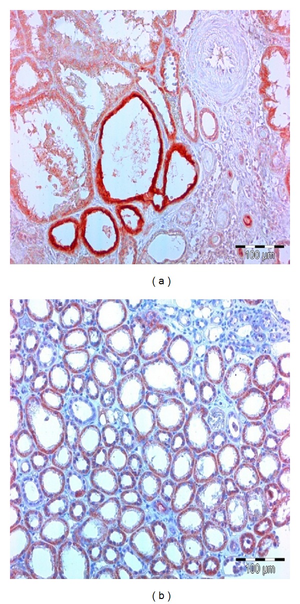
Materials and methods: Tissue KIM-1 expression was determined immunohistochemically and KIM-1 staining was scored semiquantitatively, as well as tubulointerstitialis (TIN), inflammation, atrophy, and fibrosis. Kidney function (MDRD formula) and proteinuria/day were evaluated at the time of biopsy (GFR0) and 6, 12, 24, and 36 months later.
Results: Significantly positive correlations between tissue KIM-1 expression and age (r = 0.313), TIN inflammation (r = 0.456), fibrosis (r = 0.317), and proteinuria at 6 months (r = 0.394) as well as negative correlations with GFR0 (r = -0.572), GFR6 (r = -0.442), GFR24 (r = -0.398), and GFR36 (r = -0.412) were found. Meanwhile, TIN inflammation was the best predictor of all measured kidney functions during three years, while tissue KIM-1 expression (P = 0.016) was a predictor only at 6 months after biopsy.
Conclusion: Tissue KIM-1 expression significantly predicts kidney function solely at 6 months after biopsy, when the effects of immune and nonimmune treatments are the strongest.
Figures

Similar articles
-
KIM-1 expression predicts renal outcomes in IgA nephropathy.Clin Exp Nephrol. 2013 Jun;17(3):359-64. doi: 10.1007/s10157-012-0707-2. Epub 2012 Nov 8. Clin Exp Nephrol. 2013. PMID: 23135864
-
Tubular expression of KIM-1 does not predict delayed function after transplantation.J Am Soc Nephrol. 2010 Mar;21(3):536-42. doi: 10.1681/ASN.2009040390. Epub 2009 Dec 17. J Am Soc Nephrol. 2010. PMID: 20019169 Free PMC article.
-
Hepatitis A virus cellular receptor 1/kidney injury molecule-1 is a susceptibility gene for clear cell renal cell carcinoma and hepatitis A virus cellular receptor/kidney injury molecule-1 ectodomain shedding a predictive biomarker of tumour progression.Eur J Cancer. 2013 May;49(8):2034-47. doi: 10.1016/j.ejca.2012.12.020. Epub 2013 Jan 23. Eur J Cancer. 2013. PMID: 23352434
-
Kidney injury molecule-1 in renal disease.J Pathol. 2010 Jan;220(1):7-16. doi: 10.1002/path.2642. J Pathol. 2010. PMID: 19921716 Review.
-
Biomarkers of renal function, which and when?Clin Chim Acta. 2015 Jan 1;438:350-7. doi: 10.1016/j.cca.2014.08.039. Epub 2014 Sep 3. Clin Chim Acta. 2015. PMID: 25195004 Review.
Cited by
-
Kidney injury molecule-1 expression predicts structural damage and outcome in histological acute tubular injury.Ren Fail. 2019 Nov;41(1):80-87. doi: 10.1080/0886022X.2019.1578234. Ren Fail. 2019. PMID: 30909833 Free PMC article.
-
Tissue and urinary KIM-1 relate to tumor characteristics in patients with clear renal cell carcinoma.Int Urol Nephrol. 2018 Jan;50(1):63-70. doi: 10.1007/s11255-017-1724-6. Epub 2017 Oct 19. Int Urol Nephrol. 2018. PMID: 29052086
-
Kidney Injury Molecule-1 Enhances Endocytosis of Albumin in Renal Proximal Tubular Cells.J Cell Physiol. 2016 Apr;231(4):896-907. doi: 10.1002/jcp.25181. Epub 2015 Sep 9. J Cell Physiol. 2016. PMID: 26332568 Free PMC article.
-
Increased expression of kidney injury molecule-1 and matrix metalloproteinase-3 in severe Plasmodium falciparum malaria with acute kidney injury.Int J Clin Exp Pathol. 2017 Jul 1;10(7):7856-7864. eCollection 2017. Int J Clin Exp Pathol. 2017. PMID: 31966633 Free PMC article.
-
Potential diagnostic biomarkers for chronic kidney disease of unknown etiology (CKDu) in Sri Lanka: a pilot study.BMC Nephrol. 2017 Jan 19;18(1):31. doi: 10.1186/s12882-017-0440-x. BMC Nephrol. 2017. PMID: 28103909 Free PMC article.
References
-
- Kronenberg F. Emerging risk factors and markers of chronic kidney disease progression. Nature Reviews Nephrology. 2009;5(12):677–689. - PubMed
-
- Zandi-Nejad K, Eddy AA, Glassock RJ, Brenner BM. Why is proteinuria an ominous biomarker of progressive kidney disease? Kidney International. Supplement. 2004;66(92):S76–S89. - PubMed
-
- Rodríguez-Iturbe B, Johnson RJ, Herrera-Acosta J. Tubulointerstitial damage and progression of renal failure. Kidney International. Supplement. 2005;68(99):S82–S86. - PubMed
-
- Ko GJ, Grigoryev DN, Linfert D, et al. Transcriptional analysis of kidneys during repair from AKI reveals possible roles for NGAL and KIM-1 as biomarkers of AKI-to-CKD transition. The American Journal of Physiology—Renal Physiology. 2010;298(6):F1472–F1483. - PubMed
Publication types
MeSH terms
Substances
LinkOut - more resources
Full Text Sources
Other Literature Sources
Medical
