

Arteriovenous passage times and visual field progression in normal tension glaucoma
- PMID: 24282387
- PMCID: PMC3824313
- DOI: 10.1155/2013/726912
Arteriovenous passage times and visual field progression in normal tension glaucoma
Abstract
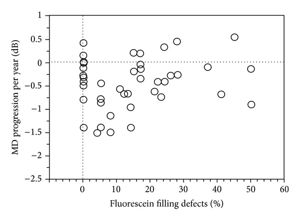
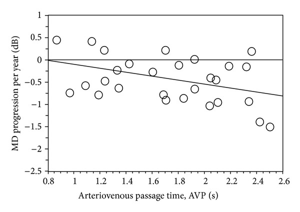
Purpose: Fluorescein angiographic studies revealed prolonged arteriovenous passage (AVP) times and increased fluorescein filling defects in normal tension glaucoma (NTG) compared to healthy controls. The purpose of this study was to correlate baseline AVP and fluorescein filling defects with visual field progression in patients with NTG.
Patients and methods: Patients with a follow-up period of at least 3 years and at least 4 visual field examinations were included in this retrospective study. Fluorescein angiography was performed at baseline using a confocal scanning laser ophthalmoscope (SLO, Rodenstock Instr.); fluorescein filling defects and AVP were measured by digital image analysis and dye dilution curves (25 Hz). Visual field progression was evaluated using regression analysis of the MD (Humphrey-Zeiss, SITA-24-2, MD progression per year (dB/year)). 72 patients with NTG were included, 44 patients in study 1 (fluorescein filling defects) and 28 patients in study 2 (AVP).
Results: In study 1 (mean follow-up 6.6 ± 1.9 years, 10 ± 5 visual field tests), MD progression per year (-0.51 ± 0.59 dB/year) was significantly correlated to the age (P = 0.04, r = -0.29) but not to fluorescein filling defects, IOP, or MD at baseline. In study 2 (mean follow-up 6.6 ± 2.2 years, 10 ± 5 visual field tests), MD progression per year (-0.45 ± 0.51 dB/year) was significantly correlated to AVP (P = 0.03, r = 0.39) but not to age, IOP, or MD at baseline.
Conclusion: Longer AVP times at baseline are correlated to visual field progression in NTG. Impaired retinal blood flow seems to be an important factor for glaucoma progression.
Figures
Similar articles
-
Prolonged retinal arteriovenous passage time is correlated to ocular perfusion pressure in normal tension glaucoma.Graefes Arch Clin Exp Ophthalmol. 2008 Aug;246(8):1147-52. doi: 10.1007/s00417-008-0807-6. Epub 2008 Apr 2. Graefes Arch Clin Exp Ophthalmol. 2008. PMID: 18386036
-
[Absolute filling defects of the optic disc in fluorescein angiograms in glaucoma--a retrospective clinical study].Klin Monbl Augenheilkd. 2001 Apr;218(4):214-21. doi: 10.1055/s-2001-14916. Klin Monbl Augenheilkd. 2001. PMID: 11392265 German.
-
Fluorescein filling defects of the optic nerve head in normal tension glaucoma, primary open-angle glaucoma, ocular hypertension and healthy controls.Ophthalmic Physiol Opt. 2006 Jan;26(1):26-32. doi: 10.1111/j.1475-1313.2005.00349.x. Ophthalmic Physiol Opt. 2006. PMID: 16390479
-
[A new approach for studying the retinal and choroidal circulation].Nippon Ganka Gakkai Zasshi. 2004 Dec;108(12):836-61; discussion 862. Nippon Ganka Gakkai Zasshi. 2004. PMID: 15656089 Review. Japanese.
-
[Aiming for zero blindness].Nippon Ganka Gakkai Zasshi. 2015 Mar;119(3):168-93; discussion 194. Nippon Ganka Gakkai Zasshi. 2015. PMID: 25854109 Review. Japanese.
Cited by
-
Ocular Blood Flow and Normal Tension Glaucoma.Biomed Res Int. 2015;2015:308505. doi: 10.1155/2015/308505. Epub 2015 Oct 19. Biomed Res Int. 2015. PMID: 26558263 Free PMC article. Review.
References
-
- Boland MV, Quigley HA. Risk factors and open-angle glaucoma: classification and application. Journal of Glaucoma. 2007;16(4):406–418. - PubMed
-
- Coleman AL, Miglior S. Risk factors for glaucoma onset and progression. Survey of Ophthalmology. 2008;53(6):3–10. - PubMed
-
- Friedman DS, Wilson MR, Liebmann JM, Fechtner RD, Weinreb RN. An evidence-based assessment of risk factors for the progression of ocular hypertension and glaucoma. American Journal of Ophthalmology. 2004;138(3):19–31. - PubMed
-
- Leske MC, Heijl A, Hussein M, Bengtsson B, Hyman L, Komaroff E. Factors for glaucoma progression and the effect of treatment: the early manifest glaucoma trial. Archives of Ophthalmology. 2003;121(1):48–56. - PubMed
-
- Nouri-Mahdavi K, Hoffman D, Coleman AL, et al. Predictive factors for glaucomatous visual field progression in the Advanced Glaucoma Intervention study. Ophthalmology. 2004;111(9):1627–1635. - PubMed
MeSH terms
LinkOut - more resources
Full Text Sources
Other Literature Sources
Medical
Miscellaneous
