. 2013 Oct 7;8(10):e76940.
doi: 10.1371/journal.pone.0076940. eCollection 2013.

Cervicothoracic multisegmental transpinal evoked potentials in humans

Affiliations

Cervicothoracic multisegmental transpinal evoked potentials in humans

Jonathan Einhorn et al. PLoS One. .

Abstract

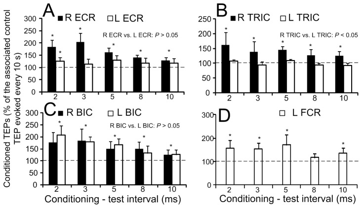
The objectives of this study were to establish the neurophysiological properties of the transpinal evoked potentials (TEPs) following transcutaneous electric stimulation of the spine (tsESS) over the cervicothoracic region, changes in the amplitude of the TEPs preceded by median nerve stimulation at group I threshold, and the effects of tsESS on the flexor carpi radialis (FCR) H-reflex in thirteen healthy human subjects while seated. Two re-usable self-adhering electrodes, connected to function as one electrode (cathode), were placed bilaterally on the clavicles. A re-usable electrode (anode) was placed on the cervicothoracic region covering from Cervical 4-Thoracic 2 and held under constant pressure throughout the experiment. TEPs were recorded bilaterally from major arm muscles with subjects seated at stimulation frequencies of 1.0, 0.5, 0.33, 0.2, 0.125, and 0.1 Hz, and upon double tsESS pulses delivered at an inter-stimulus interval of 40 ms. TEPs from the arm muscles were also recorded following median nerve stimulation at the conditioning-test (C-T) intervals of 2, 3, 5, 8, and 10 ms. The FCR H-reflex was evoked and recorded according to conventional methods following double median nerve pulses at 40 ms, and was also conditioned by tsESS at C-T intervals that ranged from -10 to +50 ms. The arm TEPs amplitude was not decreased at low-stimulation frequencies and upon double tsESS pulses in all but one subject. Ipsilateral and contralateral arm TEPs were facilitated following ipsilateral median nerve stimulation, while the FCR H-reflex was depressed by double pulses and following tsESS at short and long C-T intervals. Non-invasive transpinal stimulation can be used as a therapeutic modality to decrease spinal reflex hyper-excitability in neurological disorders and when combined with peripheral nerve stimulation to potentiate spinal output.


Conflict of interest statement

Competing Interests: The authors have declared that no competing interests exist.

Figures

Figure 1
Figure 1. Latency calculation technique.
Full-wave rectified waveform average (A) and associated cumulative sum (CUSUM) (B) characteristics of the TEPs (n = 10) elicited following non-invasive cervicothoracic transpinal stimulation and recorded from the right FCR muscle using surface EMG electrodes from one subject while at rest. The CUSUM latency, which represents the TEP latency, was determined based on the turning point of the CUSUM after the stimulus artifact.
Figure 2
Figure 2. Waveforms of transpinal evoked potentials (TEPs).
Representative examples of non-rectified waveform averages (n = 10, elicited at 0.2 Hz) of TEPs recorded following transcutaneous electric stimulation of the cervicothoracic region from the flexor carpi radialis (FCR), biceps brachii (BIC), extensor carpi radialis (ECR), and triceps brachii (TRIC) muscles in both arms.
Figure 3
Figure 3. Latency of transpinal evoked potentials (TEPs).
A, B: Non-rectified waveform averages (evoked at 0.2 Hz) of flexor carpi radialis (FCR) H-reflexes and FCR TEPs from two subjects. Note that in these subjects that the FCR TEP appeared nearly at a half latency compared to the FCR H-reflex latency. C: Overall mean latency of the FCR H-reflex and TEPs recorded from the left and right arm muscles following transcutaneous electric stimulation of the spine over the cervicothoracic region. Error bars represent the SD.
Figure 4
Figure 4. Susceptibility of transpinal evoked potentials (TEPs) to homosynaptic depression.
The overall mean amplitude of the TEPs recorded bilaterally from the right (R) and left (L) flexor carpi radialis (FCR), extensor carpi radialis (ECR), biceps brachii (BIC), and triceps brachii (TRIC) muscles for subjects 1-12 (A) and for subject 13 (B). TEPs recorded at 1.0, 0.5, 0.33, 0.2, and 0.125 Hz are presented as a percentage of the mean amplitude of the associated TEPs recorded at 0.1 Hz. Asterisks indicate statistically significant differences from control TEPs values. Error bars represent the SEM.
Figure 5
Figure 5. Transpinal evoked potentials (TEPs) and FCR H-reflexes upon double stimuli.
A, B: Non-rectified waveform averages of the FCR H-reflex and the FCR TEPs when a double pulse at an inter-stimulus interval of 40 ms was delivered to the cervicothoracic region or to the median nerve, respectively for two subjects. C: Overall average amplitude of the FCR H-reflex, FCR M-wave, and TEPs for subjects 1-12 evoked by a second pulse delivered to the median nerve or to the cervicothoracic region. D: Overall average amplitude of the FCR H-reflex, FCR M-wave, and TEPs evoked by a second pulse delivered to the median nerve or to the cervicothoracic region for subject 13. In both C and D graphs, the FCR H-reflex, FCR M-wave and TEPs were normalized to the associated potentials evoked by the first pulse. Asterisks indicate statistically significant differences from control values. Error bars indicate the SEM.
Figure 6
Figure 6. Effects of median nerve stimulation on the amplitude of transpinal evoked potentials (TEPs).
Overall mean amplitude of the TEPs recorded bilaterally from the right (R) and left (L) extensor carpi radialis (ECR), triceps brachii (TRIC), biceps brachii (BIC), and left flexor carpi radialis (FCR) muscles following median nerve stimulation at low intensities (Ia afferent). On the abscissa the conditioning-test intervals (ms) tested are indicated. Asterisks indicate statistically significant differences of conditioned TEPs from control values. Error bars indicate the SEM.
Figure 7
Figure 7. Effects of non-invasive cervicothoracic transpinal on FCR H-reflexes.
A: Non-rectified waveform averages of the FCR H-reflex in two subjects under control conditions and following transcutaneous electric stimulation of the spine (tsESS) at the conditioning-test interval of 2 ms. Note that the FCR H-reflex depression occurs with stable M-waves. B: Overall average amplitude of the FCR H-reflex conditioned by tsESS as a percentage of the control H-reflex. Asterisks indicate statistically significant differences between the conditioned and the control FCR H-reflex. C: Overall average amplitude of the FCR M-wave. Error bars indicate the SEM.

References

    1. Mills KR, Murray NMF (1986) Electrical stimulation over the human vertebral column: which neural elements are excited? Electroencephalogr Clin Neurophysiol 63: 582-589. doi: 10.1016/0013-4694(86)90145-8. PubMed: 2422007. - DOI - PubMed
    1. Maertens de Noordhout AM, Rothwell JC, Thompson PD, Day BL, Marsden CD (1988) Percutaneous electrical stimulation of lumbosacral roots in man. J Neurol Neurosurg Psychiatry 51: 174-181. doi: 10.1136/jnnp.51.2.174. PubMed: 2831302. - DOI - PMC - PubMed
    1. Cros D, Chiappa KH, Gominak S, Fang J, Santamaria J, et al. (1990) Cervical magnetic stimulation. Neurology 40: 1751-1756. doi: 10.1212/WNL.40.11.1751. PubMed: 2234432. - DOI - PubMed
    1. Chokroverty S, Picone MA, Chokroverty M (1991) Percutaneous magnetic coil stimulation of human cervical vertebral column: site of stimulation and clinical application. Electroencephalogr Clin Neurophysiol 81: 359-365. doi: 10.1016/0168-5597(91)90025-S. PubMed: 1718722. - DOI - PubMed
    1. Hunter JP, Ashby P (1994) Segmental effects of epidural spinal cord stimulation in humans. J Physiol Lond 474: 407-419. PubMed: 8014902. - PMC - PubMed

Publication types