. 2013 Nov 25;8(11):e80266.
doi: 10.1371/journal.pone.0080266. eCollection 2013.

miR-127 regulates cell proliferation and senescence by targeting BCL6

Affiliations

miR-127 regulates cell proliferation and senescence by targeting BCL6

Jingwen Chen et al. PLoS One. .

Abstract

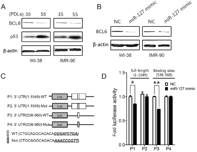
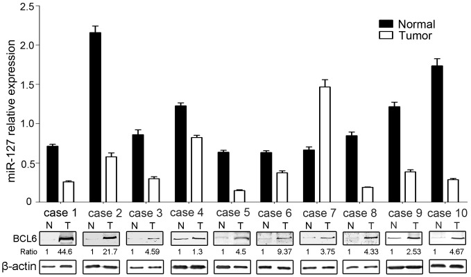
Cellular senescence occurs as a response to extracellular and intracellular stresses and contributes to aging and age-related pathologies. Emerging evidence suggests that cellular senescence also acts as a potent tumor suppression mechanism that prevents the oncogenic transformation of primary human cells. Recent reports have indicated that miRNAsact as key modulators of cellular senescence by targeting critical regulators of the senescence pathways. We previously reported that miR-127 is up-regulated in senescent fibroblasts. In this report, we identified miR-127 as a novel regulator of cellular senescence that directly targets BCL6. We further showed that miR-127 is down-regulated in breast cancer tissues and that this down-regulation is associated with up-regulation of BCL6. Over-expression of miR-127 or depletion of BCL6 inhibits breast cancer cell proliferation. Our data suggest that miR-127 may function as a tumor suppressor that modulates the oncogene BCL6.


Conflict of interest statement

Competing Interests: The authors have declared that no competing interests exist.

Figures

Figure 1
Figure 1. miR-127 is up-regulated in senescent human fibroblasts and mediates cellular senescence.
(A) Relative levels of miR-127 expression analyzed by stem-loop qRT-PCR in different PDLs of WI-38 and IMR-90 cells. U6 RNA was used for normalization. Data are presented as the mean ± SD from three independent experiments. (B) Growth curves of WI-38 and IMR-90 cells transfected with 50 nM miR-127 mimic and negative control. Growth curves were generated by cell number at the indicated times. Data are presented as the mean ± SD from three independent experiments. (C) SA-β-gal staining of senescent cells induced by miR-127. The WI-38 and IMR-90 cellswere stained for SA-β-gal activity 7 days after transfection with 50 nM miR-127 mimic or negative control. (D) Western blot analysis of cyclin D1, p53, and p21 in WI-38 and IMR-90 cells at 72 hours post-transfection with the 50 nM miR-127-3p mimic or negative control. β-actin was used for normalization. (E) Cell cycle analysis was performed at 48 h after transfection. The histogram displays the percentage changes of G0/G1 and G2/M when WI-38 cells transfected with miR-127 mimics and negative control. M1 and M2 show the spike of G0/G1 and G2/M, respectively. Data are presented as the mean ± SD from three independent experiments (*P<0.05, **P<0.01).
Figure 2
Figure 2. Stable expression miR-127 affects senescence phenotypes in WI-38 fibroblasts.
(A) Stable expression of miR-127 in WI-38 cells infected by lentiviral constructs and endogenous expression of miR-127 in senescent WI-38 cells were evaluated by qRT-PCR analysis. Data are presented as the mean ± SD from three independent experiments. (B) Growth curves of WI-38 cells stably infected with pLenti6/V5-D-premiR-127 or pLenti6/V5-D-YFP. Cells were counted every two days for two weeks. Data are presented as the mean ± SD from three independent experiments. (C) SA-β-gal staining of senescent cells induced by stable overexpression of miR-127. The WI-38 cells were stained for SA-β-gal activity 10 days after lentiviral infection. (D) Western blot analysis of senescence-associated proteins in WI-38 cells 6 days after lentiviral infection. β-actin was used for normalization.
Figure 3
Figure 3. BCL6 is a direct target of miR-127.
(A) Expression levels of BCL6 in different PDLs of WI-38 and IMR-90 cells. β-actin was used for normalization. (B) Representative Western blot analysis of BCL6 in WI-38 and IMR-90 cells transfected with 50 nM miR-127 mimic or negative control at 72 hours post-transfection. (C) Schematic representation of the luciferase reporter constructs containing full-length BCL6 3′-UTR (P1 contains wild-type miR-127 target sites and P2 is mutated in the miR-127 target sites) or the wild-type (P3) or mutant (P4) miR-127 target sites of BCL6 3′-UTR. (D) Luciferase reporter constructs (P1, P2, P3, and P4) along with pCMV-β-gal plasmid were cotransfected with miR-127 or negative control vector into MCF7 cells. Luciferase reporter assays were performed at 48 hours post-transfection. Data are presented as the mean ± SD from three independent experiments (*P<0.05, **P<0.01).
Figure 4
Figure 4. BCL6 is potentially involved in miR-127-mediated cellular senescence.
(A–B) Growth curves of WI-38 and IMR-90 cells transiently transfected with 50 nM BCL6 siRNA or negative control. Growth curves were generated by cell number at indicated times. Data are presented as the mean ± SD from three-independent experiments. (C) SA-β-gal staining of senescent cells induced by transient transfection of BCL6 siRNA. The WI-38 and IMR-90 cells were stained 7 days after transfection with 50 nM BCL6 siRNA or negative control. (D) Western blot analysis of BCL6, cyclin D1, p53, and p21 in WI-38 and IMR-90 cells transiently transfected with 50 nM BCL6 siRNA or negative control. β-actin was used for normalization. (E) Western blot analysis of the expression of BCL6 in miR-127-expressing WI-38 cells transiently transfected with pcDNA3.0 and pcDNA3.0-BCL6. (F and G) Growth curve (F) and SA-β-gal activity (G) in miR-127-expressing WI-38 cells transfected by pcDNA3.0 and pcDNA3.0-BCL6. Cells were counted every two days for a week. The cells were stained SA-β-gal activity at 7 days after transfection with pcDNA3.0 or pcDNA3.0-BCL6. Data are presented as the mean ± SD from three independent experiments (*P<0.05, **P<0.01).
Figure 5
Figure 5. MiR-127 and BCL6 modulate the proliferation of breast cancer cell lines.
(A) qRT-PCR analysis of miR-127 expression in WI-38, IMR-90, MCF7, and MDA-MB-231. U6 RNA was used for normalization. Data are presented as the mean ± SD from three independent experiments. (B) Western blot analysis of BCL6 in human fibroblasts, WI-38 and IMR-90, and the breast cancer cell lines MCF7 and MDA-MB-231. β-actin was used for normalization. (C) Growth curves of MCF7 and MDA-MB-231 cells transiently transfected with miR-127 mimic or negative control. Growth curves were generated by cell number at indicated times. Data are presented as the mean ± SD from three independent experiments. (D) Western blot analysis of BCL6, cyclin D1 and Phospho-Rb (Ser780) in MCF7 and MDA-MB-231 cells transiently transfected with the 50 nM miR-127-3p mimic or negative control. β-actin was used for normalization. (E) Cell cycle analysis was performed at 48 h after transfection.The histogram displays the percentage changes of G0/G1 and G2/M when MCF7 cells transfected with miR-127 mimics and negative control. M1 and M2 show the spike of G0/G1 and G2/M, respectively. (F) Growth curves of MCF7 and MDA-MB-231 cells transiently transfected by 50 nM BCL6 siRNA or negative control. (G) Western blot analysis of BCL6, cyclin D1 and Phospho-Rb (Ser780) in MCF7 and MDA-MB-231 cells 72 hours after transfection with 50 nM BCL6 siRNA or negative control. β-actin was used for normalization.
Figure 6
Figure 6. Expression levels of miR-127 and BCL6 in normal breast tissues and primary breast tumors.
qRT-PCR analysis of miR-127 and Western blot analysis of BCL6 in matched sets of normal tissues and primary tumors of breast. U6 RNA was used for qRT-PCR normalization. Data are presented as the mean ± SD from three independent experiments. β-actin was used for Western blot normalization. N, normal tissues; T, corresponding tumors.

References

    1. Hayflick L, Moorhead PS (1961) The serial cultivation of human diploid cell strains. Exp Cell Res 25: 585–621. - PubMed
    1. Campisi J, d’Adda di Fagagna F (2007) Cellular senescence: when bad things happen to good cells. Nat Rev Mol Cell Biol 8: 729–740. - PubMed
    1. Itahana K, Campisi J, Dimri GP (2004) Mechanisms of cellular senescence in human and mouse cells. Biogerontology 5: 1–10. - PubMed
    1. Campisi J (2013) Aging, cellular senescence, and cancer. Annu Rev Physiol 75: 685–705. - PMC - PubMed
    1. Campisi J (2005) Senescent cells, tumor suppression, and organismal aging: good citizens, bad neighbors. Cell 120: 513–522. - PubMed

Publication types

LinkOut - more resources