. 2013 Nov 25;8(11):e81741.
doi: 10.1371/journal.pone.0081741. eCollection 2013.

Higher nucleoporin-Importinβ affinity at the nuclear basket increases nucleocytoplasmic import

Affiliations

Higher nucleoporin-Importinβ affinity at the nuclear basket increases nucleocytoplasmic import

Mohammad Azimi et al. PLoS One. .

Abstract

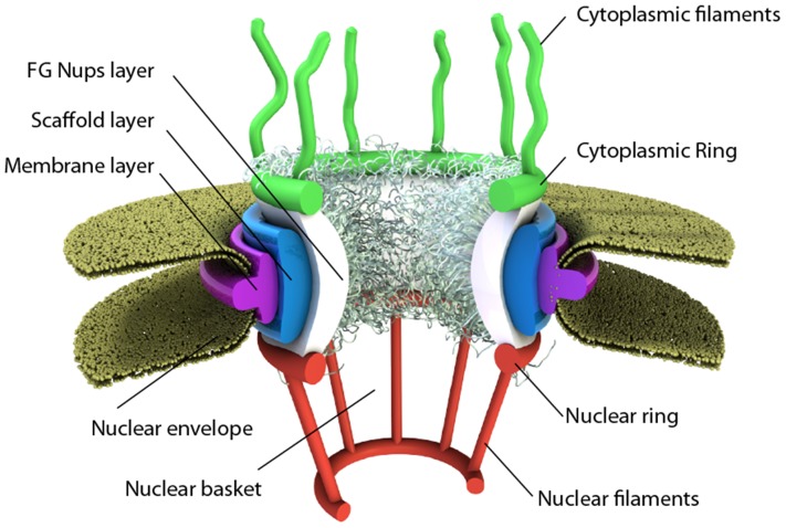
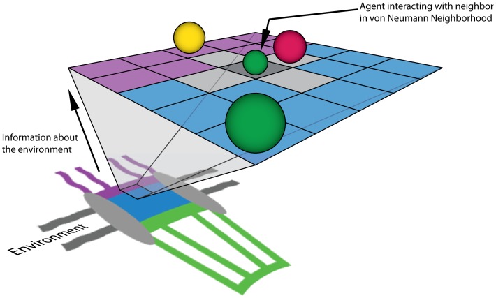
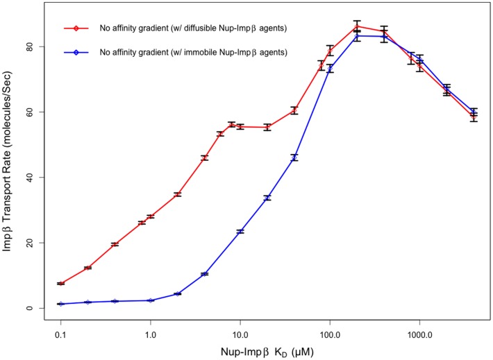
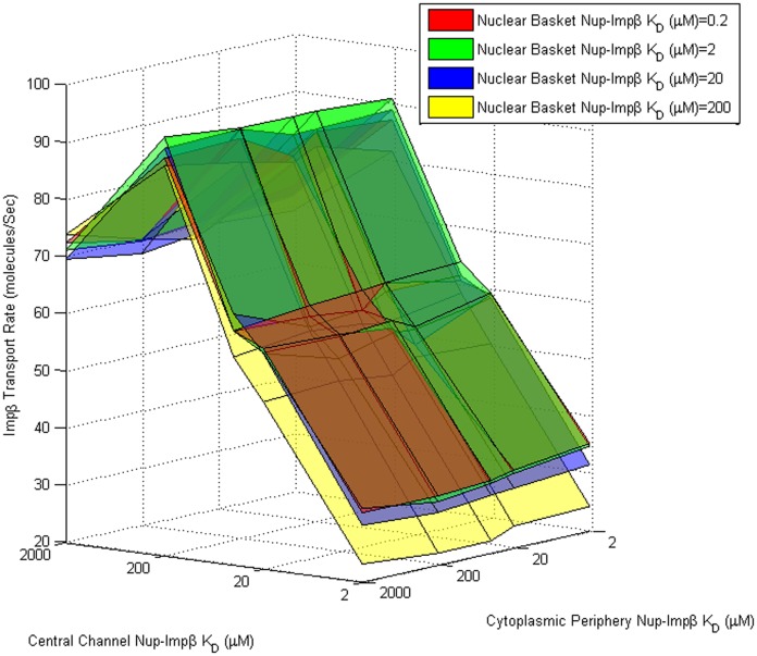
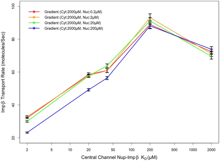
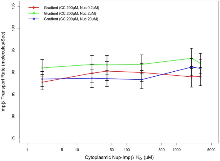
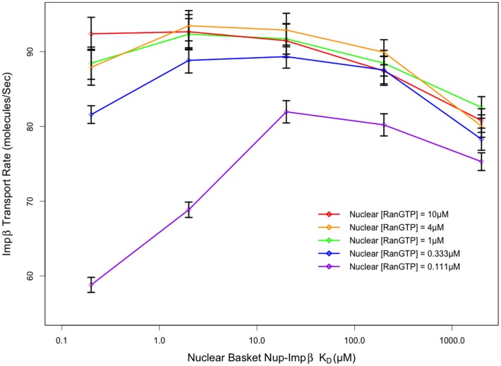
Several in vitro studies have shown the presence of an affinity gradient in nuclear pore complex proteins for the import receptor Importinβ, at least partially contributing to nucleocytoplasmic transport, while others have historically argued against the presence of such a gradient. Nonetheless, the existence of an affinity gradient has remained an uncharacterized contributing factor. To shed light on the affinity gradient theory and better characterize how the existence of such an affinity gradient between the nuclear pore and the import receptor may influence the nucleocytoplasmic traffic, we have developed a general-purpose agent based modeling (ABM) framework that features a new method for relating rate constants to molecular binding and unbinding probabilities, and used our ABM approach to quantify the effects of a wide range of forward and reverse nucleoporin-Importinβ affinity gradients. Our results indicate that transport through the nuclear pore complex is maximized with an effective macroscopic affinity gradient of 2000 µM, 200 µM and 10 µM in the cytoplasmic, central channel and nuclear basket respectively. The transport rate at this gradient is approximately 10% higher than the transport rate for a comparable pore lacking any affinity gradient, which has a peak transport rate when all nucleoporins have an affinity of 200 µM for Importinβ. Furthermore, this optimal ratio of affinity gradients is representative of the ratio of affinities reported for the yeast nuclear pore complex--suggesting that the affinity gradient seen in vitro is highly optimized.


Conflict of interest statement

Competing Interests: The authors have declared that no competing interests exist.

Figures

Figure 1
Figure 1. Schematic of the nuclear pore complex.
The pore is anchored to the nuclear envelope by a membrane layer that surrounds the scaffold layer. This scaffold layer provides structure and serves as an anchor for Nups that contain both structured domains as well as highly unstructured domains that are thought to form a barrier that excludes non-interacting molecules while allowing for selective transport of others. This central channel exhibits eight-fold rotational symmetry and has eight cytoplasmic filaments as well as eight nuclear filaments protruding into the cytoplasm and nucleoplasm respectively. The nuclear filaments are bound via a ring, resulting in a basket structure.
Figure 2
Figure 2. Simplified representation of the agent based model.
Abstract cartoon representation of the nuclear pore structure environment (not to scale) projected onto a simplified, 2-dimensional, on-lattice ABM with agents representing proteins that move within the system and interact with other agents within their von-Neumann neighborhood. The actual model consists of a three-dimensional representation of the NPC structure and physiologically relevant concentrations of biochemical factors and channel dimensions. In our model, the purple region representing the cytoplasmic periphery is treated as a compartmentalized volume containing non-interacting Nup and Impβ-interacting FG-Nup agents. Similarly, central channel (blue) and nuclear basket (green) regions are represented by compartmentalized volumes, containing both non-interacting and interacting Nup agents at physiologically meaningful concentrations. Grey regions of the diagram represent the scaffold and nuclear envelope regions of the model that are impermeable to diffusing species.
Figure 3
Figure 3. Comparison of ABM and ODE time course data.
Comparison of time-course data from an agent based model of molecular binding to that of the numerical solution of the ordinary differential equation for the same event. Probability selection using the relationship in Eq. 11 produces similar behavior to that of the numerical solution in a well-mixed system at multiple rate constants with the addition of stochasticity that is expected from natural systems.
Figure 4
Figure 4. The agent based model recapitulates the experimentally observed size-dependent permeabilities of passive cargos through the nuclear pore.
Following a simulated microinjection of non-interacting species in the cytoplasm, the in silico pore is observed to inhibit the influx of larger species while allowing smaller species to diffuse through the pore. This is in agreement with previous experimental observations.
Figure 5
Figure 5. Impβ transport rate through a pore with Nups of uniform affinity.
Transport rates for ABM simulations of Impβ through a nuclear pore containing Nups with uniform affinity (no gradient). Nup-Impβ affinity is varied from 100 nM to 4 mM. The transport rate exhibits biphasic behavior as a function of affinity. At very high affinities (low KD), Impβ is tightly bound to Nups, resulting in slow transport rates as the Nups become saturated. At very low affinities, Impβ isn't able to bind Nups as efficiently, reducing its resident time at the pore periphery and subsequently excluding it from the pore interior as a result of steric effects. Peak transport of 86.24±1.68 transports per second were observed at a Nup-Impβ affinity of 200 µM. Pores containing Nup bound Impβ agents that are capable of diffusing locally exhibit increased transport rate compared to simulation configurations where Impβ becomes immobile once bound to an FG-Nup.
Figure 6
Figure 6. Transport rate as a function of cytoplasmic, central channel, and nuclear basket Nup-Impβ affinity.
Impβ transport rate (z-axis) as a function of cytoplasmic (x-axis) and central channel (y-axis) Nup-Impβ affinity ranging from 2 µM to 2 mM. The four three-dimensional surfaces represent a range of nuclear basket affinities ranging from 0.2 µM to 200 µM. Transport rates appear to be least sensitive to cytoplasmic affinities and most sensitive to central channel and nuclear basket affinities. Varying central channel affinities results in biphasic behavior with maximum transport at KD≈200 µM. Transport rates appear to increase as nuclear basket affinity is increased up to KD≈10 µM and don't appear to show significant increase at higher affinities.
Figure 7
Figure 7. Transport rate as a function of central channel affinity.
Impβ transport rate appears most sensitive to central channel affinity, regardless of nuclear basket affinity, with a peak transport rate when Nup-Impβ affinities are on the order of 100 µM. (Cyt: Cytoplasmic periphery, Nuc: Nuclear basket)
Figure 8
Figure 8. Transport rate as a function of cytoplasmic affinity.
Transport rate appears insensitive to cytoplasmic Nup-Impβ affinity as opposed to central channel and nuclear basket Nup-Impβ affinity. An increase or decrease to affinity in the cytoplasmic region by an order of magnitude results in a change in transport rate that is within a standard error. (CC: Central Channel, Nuc: Nuclear basket)
Figure 9
Figure 9. Transport rate as a function of nuclear basket affinity.
Transport rates are very sensitive to nuclear basket Nup-Impβ affinity, with maximum transport rates emerging in the presence of a high affinity target for Impβ in the nuclear basket. Transport rates peak at an affinity of ∼2 µM with a slight decrease in transport rate as affinities are increased beyond that. This peak in transport rate doesn't appear to be due to a lack of RanGTP to terminate transport at the nuclear periphery of the pore since there aren't significant changes to transport rate under very high nuclear RanGTP concentrations. Conversely, when nuclear RanGTP concentrations are much lower than physiological values, the effect on transport rate is more noticeable.

References

    1. Reichelt R, Holzenburg A, Buhle EL Jr, Jarnik M, Engel A, et al. (1990) Correlation between structure and mass distribution of the nuclear pore complex and of distinct pore complex components. J Cell Biol 110: 883–894. - PMC - PubMed
    1. Rout MP, Aitchison JD, Suprapto A, Hjertaas K, Zhao Y, et al. (2000) The yeast nuclear pore complex: composition, architecture, and transport mechanism. J Cell Biol 148: 635–651. - PMC - PubMed
    1. Rout MP, Blobel G (1993) Isolation of the yeast nuclear pore complex. J Cell Biol 123: 771–783. - PMC - PubMed
    1. Loschberger A, van de Linde S, Dabauvalle MC, Rieger B, Heilemann M, et al. (2012) Super-resolution imaging visualizes the eightfold symmetry of gp210 proteins around the nuclear pore complex and resolves the central channel with nanometer resolution. J Cell Sci 125: 570–575. - PubMed
    1. Rout MP, Aitchison JD, Magnasco MO, Chait BT (2003) Virtual gating and nuclear transport: the hole picture. Trends Cell Biol 13: 622–628. - PubMed

MeSH terms

Substances