

A peripheral blood gene expression score is associated with plaque volume and phenotype by intravascular ultrasound with radiofrequency backscatter analysis: results from the ATLANTA study
- PMID: 24282740
- PMCID: PMC3839219
- DOI: 10.3978/j.issn.2223-3652.2013.01.02
A peripheral blood gene expression score is associated with plaque volume and phenotype by intravascular ultrasound with radiofrequency backscatter analysis: results from the ATLANTA study
Abstract
Background: A composite, peripheral gene expression score based on quantitative RNA-measurements has been validated for detecting stenosis against invasive coronary X-ray angiography. IVUS/VH has been validated for quantitative measurements of coronary plaque volume and composition and has been shown to be predictive of outcomes and treatment effects. The correlation between peripheral gene expression and coronary plaque composition by intravascular ultrasound with radiofrequency backscatter (IVUS/VH) is unknown.
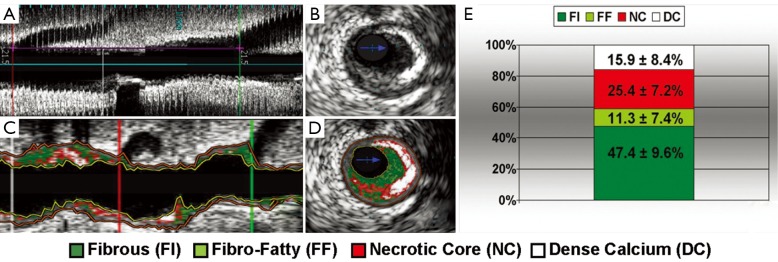
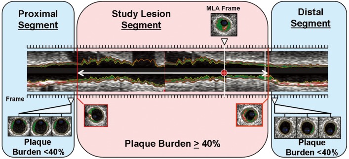
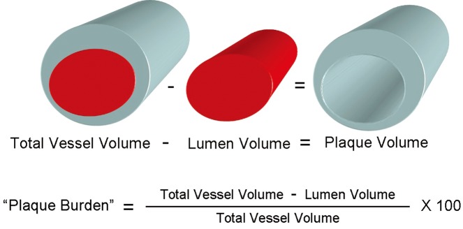
Methods: Peripheral blood gene expression score (GES) was prospectively measured in 18 patients undergoing IVUS/VH. Plaque volume and composition [fibrous tissue (FI), fibro-fatty tissue (FF), necrotic core (NC) and dense calcium (DC)] were quantified in 3 dimensions in all plaques within the entire pullback. The relationship to GES was assessed by Spearman rank correlation.
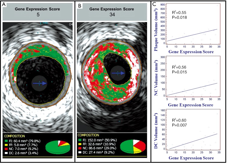
Results: Mean age was 61.1±8.6 years; 67% were male. 1,158 mm of coronary anatomy was imaged by IVUS/VH. Using a validated scale of 1-40, mean GES was 21.6±9.4. GES was associated with plaque volume (R(2)=0.55; P=0.018), NC volume (R(2)=0.56; P=0.015), DC volume (R(2)=0.60; P=0.007), and non-calcified plaque volume (R(2)=0.50; P=0.036) by Spearman rank correlation.
Conclusions: In this preliminary report, increased GES was associated with higher plaque volume and a more vulnerable plaque phenotype as evidenced by NC and DC. This composite GES is not only associated with obstructive coronary disease, but also with higher plaque volume and vulnerable phenotype.
Keywords: Gene expression; intravascular ultrasound; necrotic core; plaque volume; vulnerable plaque.
Figures
References
-
- D’Agostino RB, Sr, Vasan RS, Pencina MJ, et al. General cardiovascular risk profile for use in primary care: the Framingham Heart Study. Circulation 2008;117:743-53 - PubMed
-
- Wilson PW, D’Agostino RB, Levy D, et al. Prediction of coronary heart disease using risk factor categories. Circulation 1998;97:1837-47 - PubMed
-
- Clarke R, Peden JF, Hopewell JC, et al. Genetic variants associated with Lp(a) lipoprotein level and coronary disease. N Engl J Med 2009;361:2518-28 - PubMed
-
- Helgadottir A, Thorleifsson G, Manolescu A, et al. A common variant on chromosome 9p21 affects the risk of myocardial infarction. Science 2007;316:1491-3 - PubMed
-
- Kathiresan S, Melander O, Anevski D, et al. Polymorphisms associated with cholesterol and risk of cardiovascular events. N Engl J Med 2008;358:1240-9 - PubMed
LinkOut - more resources
Full Text Sources