. 2013 Nov 27;2(1):15.
doi: 10.1186/2046-2530-2-15.

Flagellar central pair assembly in Chlamydomonas reinhardtii

Affiliations

Flagellar central pair assembly in Chlamydomonas reinhardtii

Karl-Ferdinand Lechtreck et al. Cilia. .

Abstract

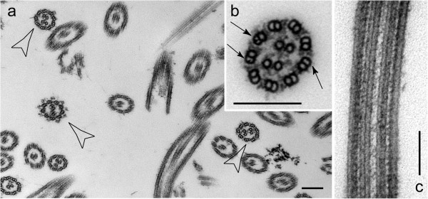
Background: Most motile cilia and flagella have nine outer doublet and two central pair (CP) microtubules. Outer doublet microtubules are continuous with the triplet microtubules of the basal body, are templated by the basal body microtubules, and grow by addition of new subunits to their distal ("plus") ends. In contrast, CP microtubules are not continuous with basal body microtubules, raising the question of how these microtubules are assembled and how their polarity is established.

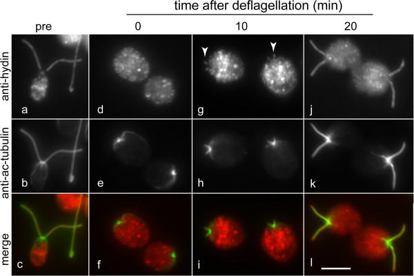
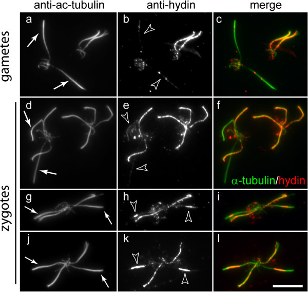
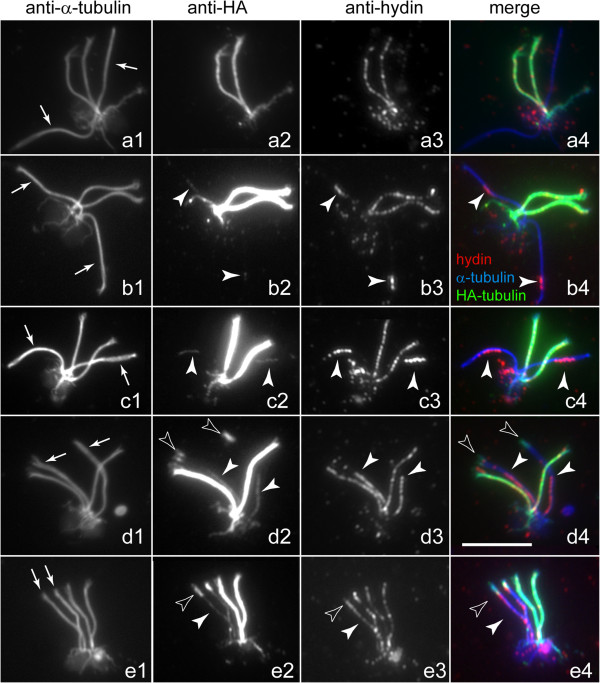
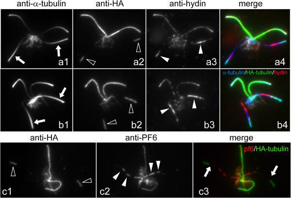
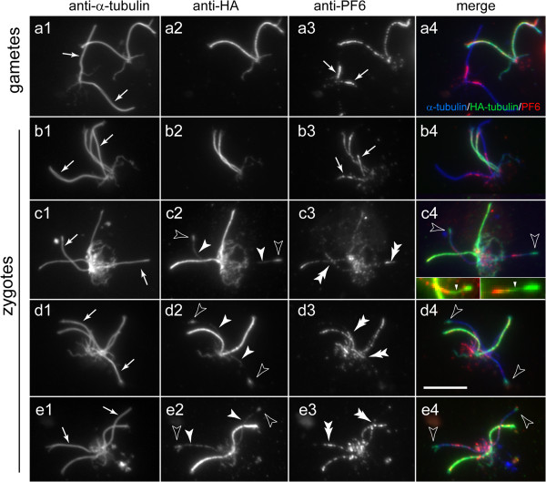
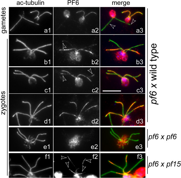
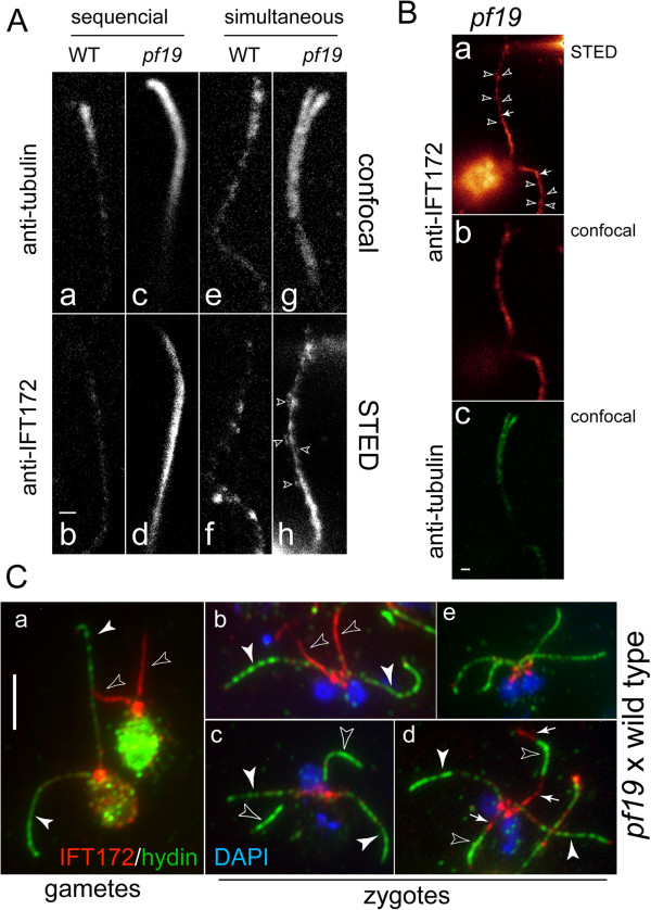
Methods: CP assembly in Chlamydomonas reinhardtii was analyzed by electron microscopy and wide-field and super-resolution immunofluorescence microscopy. To analyze CP assembly independently from flagellar assembly, the CP-deficient katanin mutants pf15 or pf19 were mated to wild-type cells. HA-tagged tubulin and the CP-specific protein hydin were used as markers to analyze de novo CP assembly inside the formerly mutant flagella.

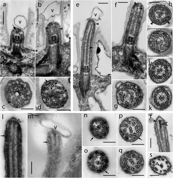
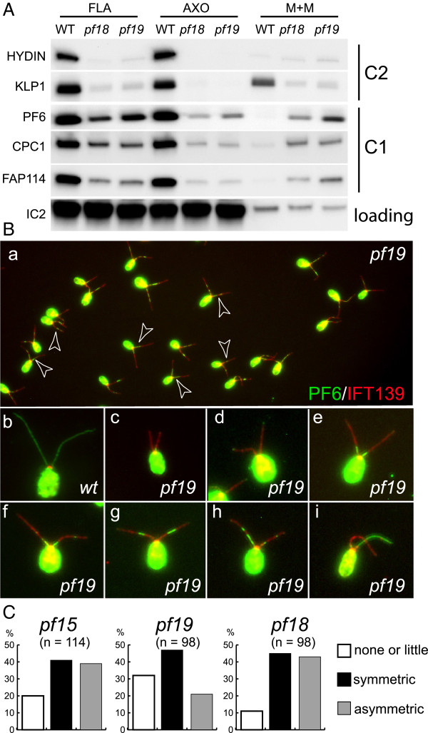
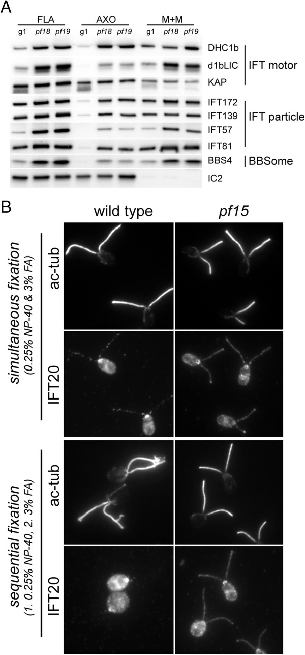
Results: In regenerating flagella, the CP and its projections assemble near the transition zone soon after the onset of outer doublet elongation. During de novo CP assembly in full-length flagella, the nascent CP was first apparent in a subdistal region of the flagellum. The developing CP replaces a fibrous core that fills the axonemal lumen of CP-deficient flagella. The fibrous core contains proteins normally associated with the C1 CP microtubule and proteins involved in intraflagellar transport (IFT). In flagella of the radial spoke-deficient mutant pf14, two pairs of CPs are frequently present with identical correct polarities.

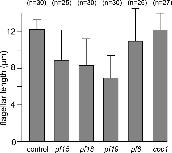
Conclusions: The temporal separation of flagellar and CP assembly in dikaryons formed by mating CP-deficient gametes to wild-type gametes revealed that the formation of the CP does not require proximity to the basal body or transition zone, or to the flagellar tip. The observations on pf14 provide further support that the CP self-assembles without a template and eliminate the possibility that CP polarity is established by interaction with axonemal radial spokes. Polarity of the developing CP may be determined by the proximal-to-distal gradient of precursor molecules. IFT proteins accumulate in flagella of CP mutants; the abnormal distribution of IFT proteins may explain why these flagella are often shorter than normal.


Figures

Figure 1
Figure 1
Ultrastructure of short regenerating flagella. Electron micrographs of cells fixed at various times after deflagellation (a-q). r, s: non-deflagellated control cells. a-d: very short regenerating flagella fixed at 7 minutes after deflagellation lack a bona fide CP. Arrows in a and d: granular material. Closed arrowheads in b: elongated microtubules. Open arrowheads in b: linear structures in the axonemal lumen which could represent a nascent CP. Arrowheads in c and d: singlet microtubules indicative of outer doublet formation. e-k: regenerating flagella at 14 minutes after amputation. Arrow in e: CP with projections. Arrowheads in e and h: fibrous material underlying the flagellar membrane. Arrowheads in f: staggered ends of the two CP microtubules. g, h: distal end of flagellum showing a ring of doublets without CP (g) and with a single CP microtubule (h). i: outer dynein arms are missing from the doublet microtubules but projections (open arrowheads) are visible on the CP. Small arrow in h and i: residual granular material in the axonemal lumen. j: projections are present on both CP microtubules (open arrowheads) but some outer dynein arms are missing (arrows). k: section revealing a full complement of dynein arms and CP projections. l-s: distal portions of regenerating flagella at 22 minutes after deflagellation (l–q) and of steady-state (r, s) flagella. Open arrowheads in l, n, o, r, and s: electron opaque tip sheet between the two CP microtubules. Arrows in l, m, n, p and o: fibrous material between the doublets and the membrane. Solid arrowhead in r: A-tubule cap forming a connection to the CP. V (in a, b, e, and m): vesicle at the flagellar tip. Bars = 200 nm (a, b, e, f, l, m, r) or 100 nm.
Figure 2
Figure 2
Hydin is incorporated early during CP assembly.WT cells before (pre) and at various time points (0, 10, 20 min) after deflagellation were analyzed by immunofluorescence microscopy using anti-hydin (a, d, g, j) and anti-acetylated tubulin (b, e, h, k). Merged images are shown in c, f, i, and l. Arrowheads in g: short flagella containing hydin. Bar = 5 μm.
Figure 3
Figure 3
Distribution of hydin during CP assembly. Gametes (a–c) and zygotes (d–l) from a mating of the CP mutant pf19 with WT (CC124) were analyzed by immunofluorescence microscopy using anti-acetylated α-tubulin and anti-hydin, as indicated. Arrows mark flagella of pf19 gametes (a) or flagella derived from pf19 in quadriflagellated zygotes (d, g, and j). Arrowheads in b and e: flagella largely lacking hydin, indicating the absence of a CP. Arrowheads in h and k: hydin accumulation in subdistal regions of flagella derived from the CP mutant. Note that the accumulation occurs symmetrically in the two flagella of a given zygote. Bar = 10 μm.
Figure 4
Figure 4
Microtubule formation during CP assembly. Zygotes obtained by mating pf15 with a WT strain expressing triple HA-tagged α-tubulin were analyzed by immunofluorescence microscopy using anti-α-tubulin (a1–e1), anti-HA (a2–e2), and anti-hydin (a3–e3). Merged images are shown in a4–e4. Arrows in a1–e1: flagella derived from pf15. Filled arrowheads: developing CP as detected with anti-HA and anti-hydin. Open arrowheads: incorporation of HA-tubulin at the distal end of flagella derived from pf15. Bar = 10 μm.
Figure 5
Figure 5
Localization of hydin in pf18 x wild type dikaryons. Zygotes from a mating of pf18 with a WT strain expressing α-tubulin fused to a triple HA-tag were analyzed by immunofluorescence microscopy using anti-α-tubulin (a1, b1), anti-HA (a2, b2), and anti-hydin (a3, b3) or anti-HA (c1) and anti-PF6 (c2). Merged images are shown in a4, b4, and c3. Arrows: flagella derived from pf18. Open arrowheads: incorporation of HA-tubulin at the distal end of flagella derived from pf18. Filled arrowheads: hydin (a3, b3) or PF6 (c2) in flagella derived from pf18. CP assembly is not apparent in flagella derived from pf18. Bar = 5 μm.
Figure 6
Figure 6
Distribution of PF6 during de novo CP assembly. Gametes and zygotes from a mating of pf15 cells with WT cells expressing triple HA-tagged α-tubulin were analyzed by immunofluorescence microscopy using anti-α-tubulin (a1–e1), anti-HA (a2–e2), and anti-PF6 (a3–e3). Merged images are shown in a4–e4. Arrows in a1–e1: flagella of pf15 gametes (a1) or zygotic flagella derived from pf15(b1–e1). Filled arrowheads in c2–e2: developing CP as detected with anti-HA. Open arrowheads in c–e: incorporation of HA-tubulin at the distal ends of flagella derived from pf15. Arrows in a3 and b3: PF6 is present in the proximal regions of flagella of pf15 gametes and of flagella derived from pf15 in early quadriflagellates. Double arrowheads: position of PF6 during CP development. Inserts in c4: PF6 (red) is only present in the proximal regions of the assembling CP; accumulation of HA-tubulin (green) at the distal tip represents tubulin turnover in the outer doublets. Arrowheads: PF6-deficient regions of the newly formed CP. Bar = 10 μm.
Figure 7
Figure 7
Distribution of PF6 during repair of pf6 mutant flagella. Gametes (a) and zygotes (b–f) from matings of pf6 with WT(a–d), pf6 with pf15(f), and, as a control for antibody specificity, pf6 with pf6(e) were labeled with antibodies to acetylated α-tubulin and PF6. Merged images, mostly counterstained with DAPI to visualize the nuclei, are shown in the third column. Arrowheads in b2 and c2: incorporation of PF6 near the tip of zygotic flagella derived from pf6. In f, note the strikingly different distribution of PF6 in the lower pair of flagella derived from the CP-deficient strain pf15 vs. the upper pair of flagella derived from the PF6-deficient strain. Bar = 10 μm.
Figure 8
Figure 8
C1-associated proteins are present in CP-deficient flagella. (A) Western blot of flagella (FLA), axonemes (AXO), and the membrane + matrix fractions (M + M) isolated from WT and the CP-deficient mutants pf18 and pf19 probed with antibodies as indicated. Only traces of the C2 proteins hydin and KLP1 were detected in pf18 and pf19 flagella and both proteins were almost completely released into the detergent extract (M + M). In contrast, significant amounts of the C1 proteins PF6, CPC1, and FAP114 were present in the CP-deficient flagella and a significant proportion of these proteins remained in the axonemal fraction following detergent extraction. The outer dynein arm intermediate chain IC2 was used as a loading control. (B) To analyze the distribution of PF6 in CP-deficient flagella, methanol-fixed vegetative WT(b) and pf19 cells (a, c–i) were labeled with antibodies to PF6 and IFT139. Arrowheads (subpanel a) mark cells with a nearly symmetrical distribution of PF6 in both flagella. Note the accumulation of IFT139 in pf19 flagella compared to WT flagella. (C) The distribution of PF6 in pf15, pf18, and pf19 flagella was scored as reduced or absent (e.g. B c, d), nearly symmetrical (e.g. B e, f, g), or asymmetric (e.g. B h, i) within the two flagella of a given cell.
Figure 9
Figure 9
IFT proteins accumulate in CP-deficient flagella. (A) Western blot probing isolated flagella (FLA), axonemes (AXO), and the membrane + matrix fraction (M + M) of WT (g1) and the CP-deficient mutants pf18 and pf19 with the antibodies indicated. Similar results were obtained for pf15 (not shown). (B) Immunofluorescence microscopy of WT and pf15 cells extracted with detergent and fixed with formaldehyde either simultaneously (top) or sequentially (bottom) and then stained with antibodies to acetylated tubulin and IFT20. Note retention of IFT20 in pf15 but not WT axonemes extracted before fixation.
Figure 10
Figure 10
IFT172 is part of the fibrous core of pf19 flagella. (A) Confocal (a, c, e, g) and STED (b, d, f, h) images of WT flagella stained with anti-β-tubulin (a, c, e, g) and anti-IFT172 (b, d, f, h). Cells were extracted and fixed either sequentially (a-d) or simultaneously (e-h). Arrowheads in h: IFT172-containing particles flanking a central rod containing IFT172; note the absence of such particles in the flagella (b and d) derived from cells extracted first with detergent. Note also that the central IFT172-containing core, present in pf19 flagella (d) but not WT flagella (b), persists after detergent extraction (h) and is absent from the distal end of the cilium (compare c and d). (B) Overview of the cell corresponding to g/h showing the IFT172 signal in STED (a) and confocal illumination (b) and the tubulin signal in confocal illumination (c). Arrowheads: IFT172-containing particles flanking the central rods (arrows), which are stained strongly by the IFT172 antibody. (C)WT and pf19 gametes (a) and the resulting zygotes (b–e) were stained with antibodies to hydin (green) and IFT172 (red); DAPI staining is shown in blue. Open arrowheads: flagella of pf19 gametes (a) or zygotic flagella derived from pf19(b–d). Closed arrowheads: flagella of WT gametes (a) or zygotic flagella derived from WT(b–d). Small arrows in d: residual IFT172 flanking the developing CP as visualized by anti-hydin. Bar = 5 μm.
Figure 11
Figure 11
Flagellar length is reduced in CP-deficient mutants. Bar graph showing average steady-state flagellar length in WT, the CP-deficient mutants pf15, pf18, and pf19, and CP mutants pf6 (lacking the C1a projection) and cpc1 (lacking the C1b projection [34]). The number of vegetative cells scored is indicated. Error bars indicate the standard deviation.
Figure 12
Figure 12
Multiple CPs in pf14 flagella. Standard transmission EM of isolated pf14 axonemes. (a) Axonemes with four CP microtubules are marked. (b, c) Axonemes with two CPs in cross and longitudinal section. When two CPs are present, both always have correct, identical polarities, as seen in the cross section. Arrows point to beak-like projections in doublets 1, 5, and 6, indicating that the section is from the proximal ~1/3 of the flagellum [31].

Similar articles

Cited by

References

    1. Mitchell DR, Sale WS. Characterization of a Chlamydomonas insertional mutant that disrupts flagellar central pair microtubule-associated structures. J Cell Biol. 1999;2(2):293–304. - PMC - PubMed
    1. Mitchell DR, Smith B. Analysis of the central pair microtubule complex in Chlamydomonas reinhardtii. Methods Cell Biol. 2009;2:197–213. - PubMed
    1. DiPetrillo CG, Smith EF. Pcdp1 is a central apparatus protein that binds Ca(2+)-calmodulin and regulates ciliary motility. J Cell Biol. 2010;2(3):601–612. - PMC - PubMed
    1. Lechtreck KF, Delmotte P, Robinson ML, Sanderson MJ, Witman GB. Mutations in hydin impair ciliary motility in mice. J Cell Biol. 2008;2(3):633–643. - PMC - PubMed
    1. Lee L, Campagna DR, Pinkus JL, Mulhern H, Wyatt TA, Sisson JH, Pavlik JA, Pinkus GS, Fleming MD. Primary ciliary dyskinesia in mice lacking the novel ciliary protein Pcdp1. Mol Cell Biol. 2008;2(3):949–957. - PMC - PubMed

LinkOut - more resources