

Giant mucocele originating from the middle concha in a 5-year-old child: a case report
- PMID: 24284013
- PMCID: PMC4219181
- DOI: 10.1186/1752-1947-7-246
Giant mucocele originating from the middle concha in a 5-year-old child: a case report
Abstract
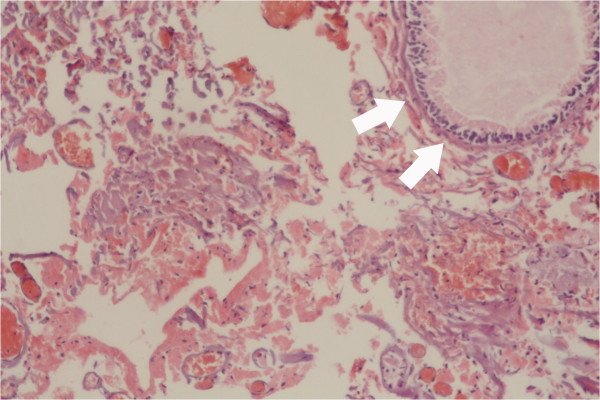
Introduction: Mucoceles are mucus-filled, epithelial-lined sacs that slowly develop in the paranasal sinuses when sinus or concha bullosa drainage is obstructed by inflammatory processes, trauma, or prior surgery. They are extremely rare in children. Symptoms usually arise from the nasal obstruction or compression of neighboring structures.
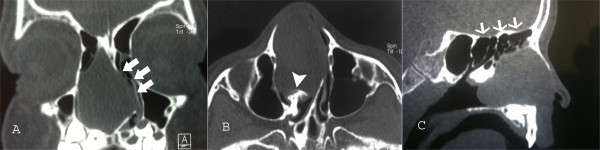
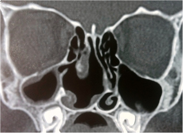
Case presentation: This case report describes a 5-year-old Turkish boy with a 3-year history of nasal obstruction. A computed tomography scan showed a well-defined soft tissue density lesion, seemingly originating in the region of the middle concha and was suggestive of a middle concha mucocele. The mass was removed by endoscopic sinus surgery.
Conclusions: In the case of a child presenting with nasal obstruction, mucocele should be remembered in the differential diagnosis of intranasal tumors. Computed tomography and magnetic resonance imaging are helpful in making the diagnosis and endoscopic nasal surgery has proven successful in the treatment.
Figures
References
-
- Loehrl TA, Leopold DA. Sphenoethmoidal mucocele presenting with bilateral visual compromise. Ann Otol Rhinol Laryngol. 2000;109:608–610. - PubMed
-
- Alvarez RJ, Liu NJ, Isaacson G. Pediatric ethmoid mucoceles in cystic fibrosis: long-term follow-up of reported cases. Ear Nose Throat J. 1997;76:538–539. 543–536. - PubMed
LinkOut - more resources
Full Text Sources
Other Literature Sources
