. 2014 Mar 1;20(5):1135-45.
doi: 10.1158/1078-0432.CCR-13-0956. Epub 2013 Nov 27.

A genome-wide aberrant RNA splicing in patients with acute myeloid leukemia identifies novel potential disease markers and therapeutic targets

Affiliations

A genome-wide aberrant RNA splicing in patients with acute myeloid leukemia identifies novel potential disease markers and therapeutic targets

Sophia Adamia et al. Clin Cancer Res. .

Abstract

Purpose: Despite new treatments, acute myeloid leukemia (AML) remains an incurable disease. More effective drug design requires an expanded view of the molecular complexity that underlies AML. Alternative splicing of RNA is used by normal cells to generate protein diversity. Growing evidence indicates that aberrant splicing of genes plays a key role in cancer. We investigated genome-wide splicing abnormalities in AML and based on these abnormalities, we aimed to identify novel potential biomarkers and therapeutic targets.

Experimental design: We used genome-wide alternative splicing screening to investigate alternative splicing abnormalities in two independent AML patient cohorts [Dana-Farber Cancer Institute (DFCI) (Boston, MA) and University Hospital de Nantes (UHN) (Nantes, France)] and normal donors. Selected splicing events were confirmed through cloning and sequencing analysis, and than validated in 193 patients with AML.

Results: Our results show that approximately 29% of expressed genes genome-wide were differentially and recurrently spliced in patients with AML compared with normal donors bone marrow CD34(+) cells. Results were reproducible in two independent AML cohorts. In both cohorts, annotation analyses indicated similar proportions of differentially spliced genes encoding several oncogenes, tumor suppressor proteins, splicing factors, and heterogeneous-nuclear-ribonucleoproteins, proteins involved in apoptosis, cell proliferation, and spliceosome assembly. Our findings are consistent with reports for other malignances and indicate that AML-specific aberrations in splicing mechanisms are a hallmark of AML pathogenesis.

Conclusions: Overall, our results suggest that aberrant splicing is a common characteristic for AML. Our findings also suggest that splice variant transcripts that are the result of splicing aberrations create novel disease markers and provide potential targets for small molecules or antibody therapeutics for this disease.


Conflict of interest statement

Disclosure of Potential Conflicts of Interest

J.D. Griffin has commercial research grant from Jenssen Pharmaceuticals and Novartis Pharmaceuticals and is a consultant/advisory board member of Curis Pharmaceuticals and Novartis Pharmaceuticals. No potential conflicts of interest were disclosed by the other authors.

Figures

Figure 1
Figure 1
A–C, aberrant splicing event frequencies detected in patients with AML; CIRCOS plots (A–C) describe distributions of the genome-wide splicing events in patients with AML. Outer circles represent the human reference genomes; spliced gene frequencies in patients are shown as dot plots, with each dot representing a single spliced gene, which is spliced in patients recurrently but is absent in normal donors. The greater the distance of a given dot from the outer circles, the more frequently the gene is spliced in patients with AML. The inner circle represents chromosomes. More than a hundred differentially and recurrently spliced genes are detected in chromosomes 1, 2, and 17 of the DFCI cohort (A), and in chromosomes 1 to 3, 11, 12, 17, and 19 of the UHN cohort (B). CIRCOS plot was generated on the basis of the list of the genes we have identified as spliced in at least 30% or more patients from DFCI cohort; 75% (262 of 348) of these spliced genes were frequently (range 2–34 patients) spliced in the UHN cohort (C). D, genome-wide splicing patterns detected in patients with AML. On the basis of probe set expression levels, a complex splicing pattern as detected in patients with AML compared with normal donors, summarized in this figure as “pattern 1”—full or partial exon(s) skipping (sequence exclusions) and “pattern 2”—full or partial cryptic exon or intron retentions (sequence inclusions). On these diagrams, dark black boxes represent exons and solid lines introns; on the exons, partial deletions are marked by the arrows, and intron retentions are shown as transparent gray boxes.
Figure 2
Figure 2
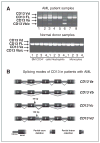
Exon array validation studies. A, CD13 full length (FL) and splice variant transcript amplifications for cloning and sequencing analyses. CD13 novel splice variant transcripts were amplified from patients with AML and normal donor samples. After cycling, RT-PCR products were subjected to 1% agarose gel electrophoresis. Bands representing different splice variant transcripts were cut out and cloned and sequenced to identify modes of splicing. B, splicing modes of CD13 identified in patients with AML. CD13 splicing pattern was identified by cloning and sequencing analyses carried out in patients with AML. Obtained CD13 novel variant sequences were aligned with the published sequence for human CD13 mRNA (gi:178535 NCBI). The CD13Va and CD13-Vb splice variants are the result of complete deletions of exon 4 (140 bp) or exons 4 (140 bp) and 6 (155 bp), respectively, whereas CD13Vc is a result of a partial deletion of exon 4 (44 bp) and a complete deletion of exon 6 (155 bp). The CD13-Vd splice variant transcript is a result of activation of a cryptic 3′ splicing site on intron 4. This splicing aberration causes the partial retention of intron 4 (132 bps). The splicing event does not lead to a frameshift, and the native stop codon is retained. However, as a result of these aberrations, 140 bp is spliced out from CD13Va, 295 bp from CD13Vb, and 199 bp from CD13Vc transcripts. Thus, all these transcripts are subjected to frameshift.
Figure 3
Figure 3
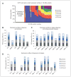
CD13 splice variants are commonly expressed in patients with AML and are associated with disease status. This figure displays overall expression patterns of CD13 full length (FL) and their splice variants transcripts. On the figure, the x-axes display 193 patient samples and the y-axes relative fluorescent units (RFU). PCR product RFU = log 2RFU. RFU is a unit of measurement calculated relative to the size standards included in each reaction. For relative level determination, product levels were kept below 3500 RFU and size standard levels were within 500 to 800 units as recommended by the manufacturer. All calculations and instrument calibration were done according to Applied Biosystems recommendations. A, CDF13 and splice variant expression profiles in 193 patients with AML. B–D, display overall expression patterns of CD13 full length and novel splice variants transcripts over the course of the disease in 14 patients. Patient samples were collected and expression profiles of CD13 full length and novel splice variant transcripts were evaluated. B, at diagnosis (Dx) and remission (REM), (C) at relapse (REL), diagnosis (Dx), and during persistent disease (PER), D) at diagnosis (Dx) and relapse (REL). A total 29 AML patient samples were collected including: ten samples obtained at diagnosis (Dx 1- Dx 3 and Dx 8 - Dx 14); three samples obtained at remission (REM 1- REM 3); six samples obtained during regular visits over the course of refractory disease (PER 4, 5, 7); nine samples obtained at relapse (REL 6, REL 8- REL 14). Samples taken at first and second relapse or visits are marked as −1 or −2.

References

    1. Ast G. How did alternative splicing evolve? Nat Rev Genet. 2004;5:773–82. - PubMed
    1. Cartegni L, Chew SL, Krainer AR. Listening to silence and understanding nonsense: exonic mutations that affect splicing. Nat Rev Genet. 2002;3:285–98. - PubMed
    1. Jurica MS, Moore MJ. Pre-mRNA splicing: awash in a sea of proteins. Mol Cell. 2003;12:5–14. - PubMed
    1. Jurica MS. Searching for a wrench to throw into the splicing machine. Nat Chem Biol. 2008;4:3–6. - PubMed
    1. Hastings ML, Krainer AR. Pre-mRNA splicing in the new millennium. Curr Opin Cell Biol. 2001;13:302–9. - PubMed

Publication types