. 2013 Dec 10;110(50):20218-23.
doi: 10.1073/pnas.1313905110. Epub 2013 Nov 27.

Inducing host protection in pneumococcal sepsis by preactivation of the Ashwell-Morell receptor

Affiliations

Inducing host protection in pneumococcal sepsis by preactivation of the Ashwell-Morell receptor

Prabhjit K Grewal et al. Proc Natl Acad Sci U S A. .

Abstract

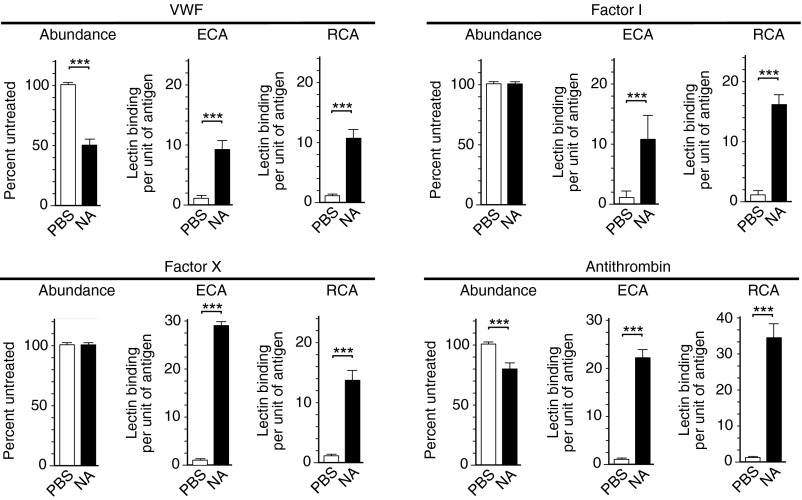
The endocytic Ashwell-Morell receptor (AMR) of hepatocytes detects pathogen remodeling of host glycoproteins by neuraminidase in the bloodstream and mitigates the lethal coagulopathy of sepsis. We have investigated the mechanism of host protection by the AMR during the onset of sepsis and in response to the desialylation of blood glycoproteins by the NanA neuraminidase of Streptococcus pneumoniae. We find that the AMR selects among potential glycoprotein ligands unmasked by microbial neuraminidase activity in pneumococcal sepsis to eliminate from blood circulation host factors that contribute to coagulation and thrombosis. This protection is attributable in large part to the rapid induction of a moderate thrombocytopenia by the AMR. We further show that neuraminidase activity in the blood can be manipulated to induce the clearance of AMR ligands including platelets, thereby preactivating a protective response in pneumococcal sepsis that moderates the severity of disseminated intravascular coagulation and enables host survival.


Conflict of interest statement

The authors declare no conflict of interest.

Figures

Fig. 1.
Fig. 1.
Intravenous neuraminidase treatment in platelet desialylation and turnover, bleeding time increases, and Gp1bα-dependent platelet clearance involving AMR function. (A) Circulating platelet abundance, (B) Erythrina cristagalli lectin (ECA) and Ricinus communis-1 agglutinin (RCA-I) lectin binding at the platelet surface, and (C) bleeding times in response to a single i.v. administration of neuraminidase (NA) or PBS in WT mice. (D) Platelet abundance and (E) bleeding times among mice identically treated but lacking either Asgr1 or Asgr2 components of the AMR. (F) Gp1bα deficiency and neuraminidase-induced desialylation among platelets of indicated genotypes. (G) Circulating platelet clearance after platelet isolation, labeling, and transfer among mice of indicated donor and recipient genotypes. (H) Platelet abundance in WT or GP1bα-deficient mice 24 h after i.v. neuraminidase or PBS treatment. Antibody to CD41 was used to detect and quantify platelets in whole blood. In these studies mice were treated with either 5 U/kg of Arthrobacter urefaciens neuraminidase or PBS control. Studies included between 12 and 24 age-matched adult mice of indicated genotypes. ***P < 0.001.
Fig. 2.
Fig. 2.
Effects of intravenous neuraminidase treatment on coagulopathy, organ damage, and host survival in SPN sepsis. (A) WT mice were infected by i.p. injection of 104 cfu of SPN isolate D39, and survival was followed over time among cohorts receiving either AUS neuraminidase (NA) (5 U/kg) or PBS at 8 h after infection. More than 40 mice receiving each treatment were analyzed from multiple independent experiments. (B) Representative macroscopic views of liver and spleen 48 h after SPN infection. (C) Serum levels of alanine aminotransferase activity. (D) Histopathological analyses of liver and spleen tissue 48 h after infection exhibited fibrin thrombi and empty vessels (open arrows) indicative of thromboembolic occlusions. Functioning blood vessels containing red blood cells are also denoted (closed arrows). Pyknotic bodies indicating cell death are marked (asterisks). Fibrin clots, pyknotic bodies, and splenic hemorrhage were quantified. Studies in B–D compared 12–24 age-matched adult mice of indicated genotypes. ***P < 0.001.
Fig. 3.
Fig. 3.
AMR function in host protection after neuraminidase treatment during SPN sepsis. (A) Survival among cohorts of mice lacking either the Asgr1 or Asgr2 component of the AMR in the presence or absence of neuraminidase (NA) or PBS delivered by i.v. injection 8 h after i.p. injection of SPN isolate D39 (104 cfu). Each cohort of mice received either AUS neuraminidase (5 U/kg) or PBS at 8 h after infection. More than 40 mice of each genotype receiving the indicated treatments from multiple independent experiments were analyzed. (B) Serum levels of alanine aminotransferase activity at 48 h. (C) Representative macroscopic views of the liver and spleen 48 h after SPN infection. (D) Histopathological analyses of liver and spleen tissue 48 h after infection indicated fibrin deposition and empty blood vessels (open arrows). Pyknotic bodies indicating cell death are marked (asterisks). Fibrin clots, pyknotic bodies, and splenic hemorrhage were quantified. At least 12 age-matched mice of each genotype were analyzed in B–D from multiple independent experiments. ***P < 0.001, **P < 0.01, *P < 0.05.
Fig. 4.
Fig. 4.
Coagulopathy and host protection after anti-CD41 treatment and platelet depletion during SPN sepsis. (A) Platelet abundance in circulation 2 h after i.v. injection of anti-CD41 antibody or polyclonal IgG at indicated doses. (B) Platelet abundance in circulation at indicated times after a single i.v. injection (0.5 mg/kg) of either anti-CD41 or IgG. (C) ECA and RCA-I lectin binding at the platelet surface 2 h after i.v. injection (0.5 mg/kg) of either anti-CD41 or IgG. (D) Bleeding times after i.v. injection (0.5 mg/kg) of either anti-CD41 or IgG. (E) Representative macroscopic and histopathologic views of liver and spleen 48 h after SPN infection. Fibrin deposition and empty blood vessels are marked (open arrows). Blood vessels containing red blood cells are also denoted (closed arrows). Areas of pyknotic bodies indicating cell death are marked (asterisks). Fibrin clots, pyknotic bodies, and splenic hemorrhage were quantified. (F) Serum alanine aminotransferase activity. (G) Survival among mice receiving 0.5 mg/kg of either anti-CD41 or IgG 8 h after i.p. injection of SPN isolate D39 (104 cfu). At least 12 age-matched mice of each genotype were analyzed in B–D from multiple independent experiments. In G, more than 40 mice of each genotype receiving each treatment were analyzed in multiple independent experiments. Findings shown are representative of results obtained using anti-CD41 and IgG in either WT or AMR-deficient mice. ***P < 0.001.
Fig. 5.
Fig. 5.
(A–D) Treatment with neuraminidase (NA), but not anti-CD41 antibody, induces a hypocoagulative state in blood plasma. aPTT and partial thrombin (PT) time were determined in plasma after either in vivo or in vitro treatments with neuraminidase or anti-CD41 in parallel comparisons with PBS or IgG control treatments.
Fig. 6.
Fig. 6.
Blood coagulation factor abundance or activity after (A) i.v. neuraminidase (NA) treatment in the presence or absence of AMR function in mice, and (B) in vitro neuraminidase treatment of human blood plasma. Wild-type or AMR-deficient mice were bled 2 h after i.v. treatment with either AUS NA (5 U/kg) or PBS. Plasma was isolated for measurements of the abundance or activity of specific blood coagulation factors, as previously described (24, 25). Measurements of glycoprotein abundance were obtained with anti-factor antibodies. Factor-specific enzyme activities were measured where indicated.
Fig. 7.
Fig. 7.
Blood coagulation factor abundance and desialylation after i.v. neuraminidase (NA) treatment. Coagulation factor protein abundance measured by antibodies was further analyzed by ECA and RCA-I lectins to detect exposed galactose following i.v. treatment with either AUS NA (5 U/kg) or PBS. Ratios of exposed galactose per unit of protein antigen were calculated as indicated and as previously described (25).

References

    1. Stearns-Kurosawa DJ, Osuchowski MF, Valentine C, Kurosawa S, Remick DG. The pathogenesis of sepsis. Annu Rev Pathol. 2011;6:19–48. - PMC - PubMed
    1. Levi M, Schultz M, van der Poll T. Sepsis and thrombosis. Semin Thromb Hemost. 2010;36:367–377. - PubMed
    1. Marshall JC. Inflammation, coagulopathy, and the pathogenesis of multiple organ dysfunction syndrome. Crit Care Med. 2001;29(7) Suppl:S99–S106. - PubMed
    1. Giamarellos-Bourboulis EJ. The failure of biologics in sepsis: Where do we stand? Int J Antimicrob Agents. 2013;42(Suppl):S45–S47. - PubMed
    1. Angus DC. The search for effective therapy for sepsis: Back to the drawing board? JAMA. 2011;306(23):2614–2615. - PubMed

Publication types

LinkOut - more resources