. 2014 Jan;141(1):148-57.
doi: 10.1242/dev.101550. Epub 2013 Nov 27.

β-catenin regulates Pax3 and Cdx2 for caudal neural tube closure and elongation

Affiliations

β-catenin regulates Pax3 and Cdx2 for caudal neural tube closure and elongation

Tianyu Zhao et al. Development. 2014 Jan.

Abstract

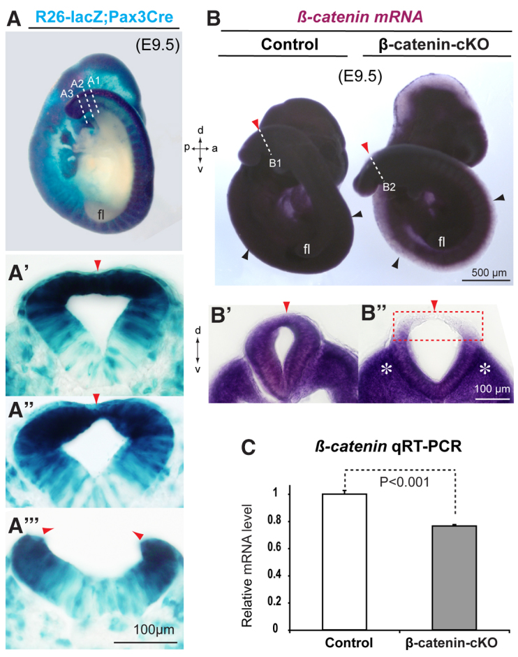
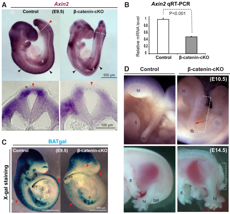
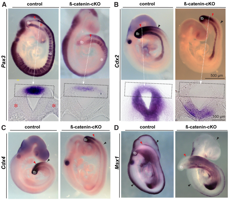
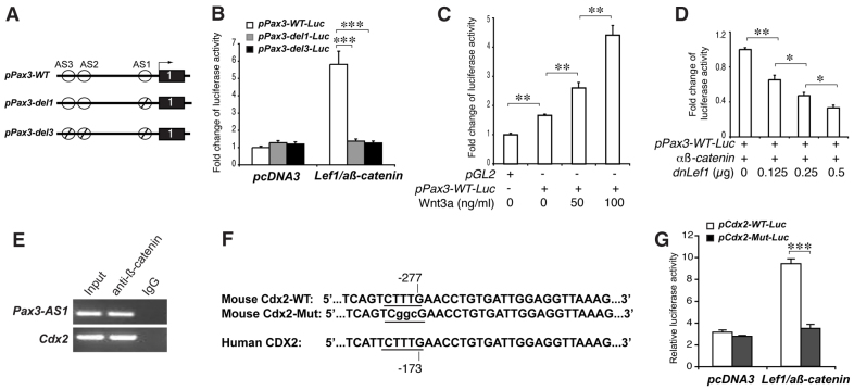
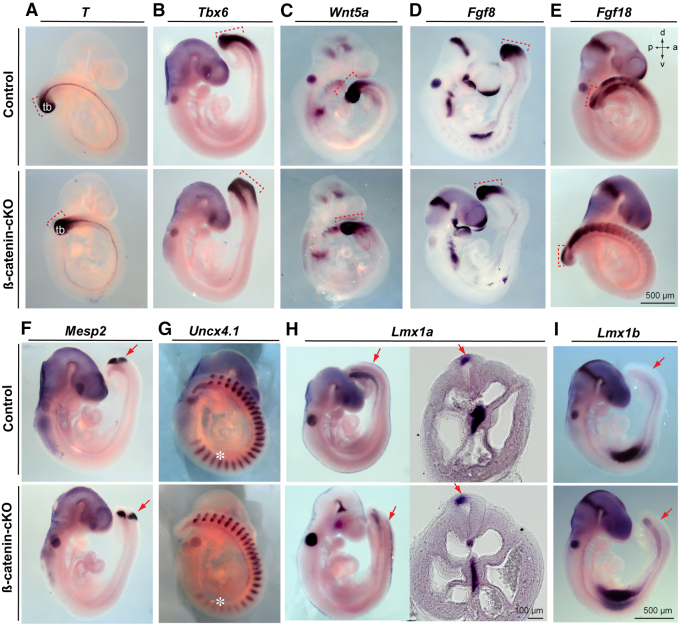
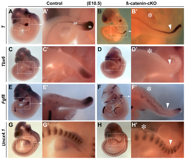
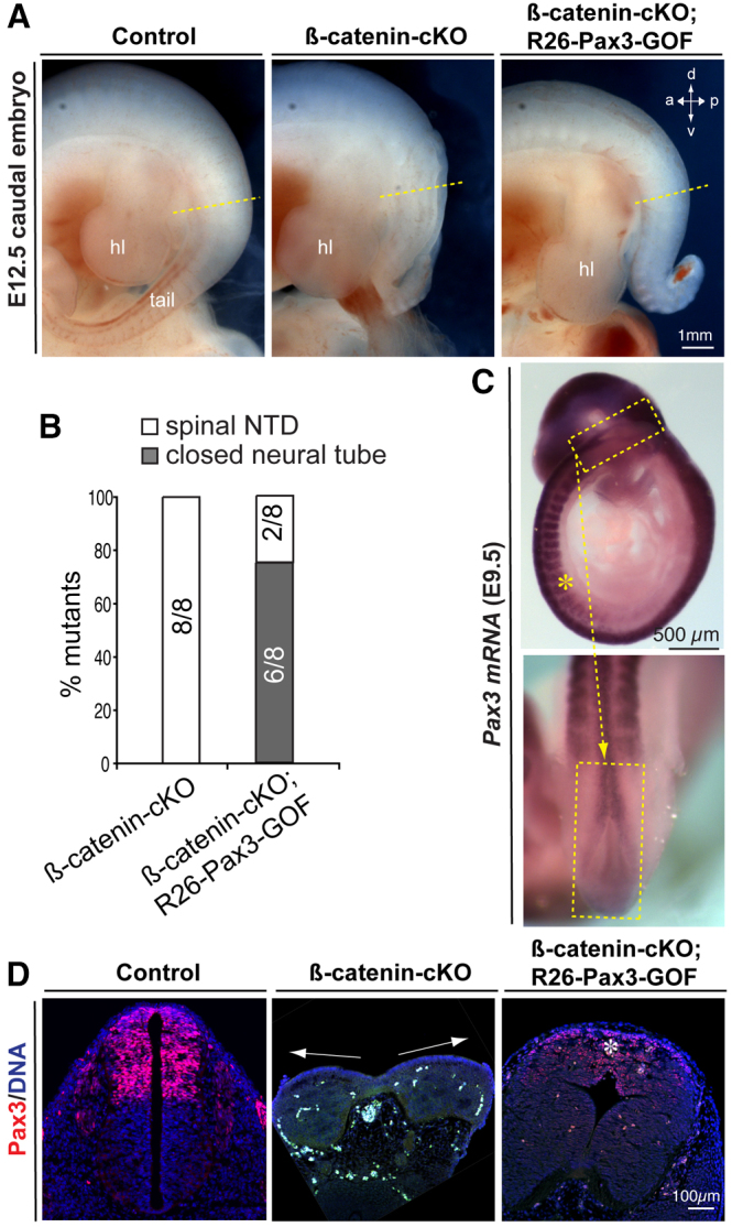
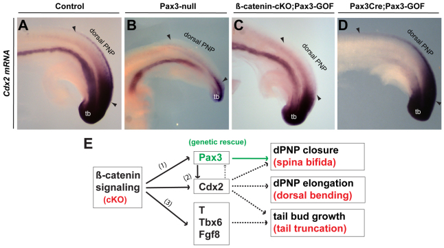
Non-canonical Wnt/planar cell polarity (PCP) signaling plays a primary role in the convergent extension that drives neural tube closure and body axis elongation. PCP signaling gene mutations cause severe neural tube defects (NTDs). However, the role of canonical Wnt/β-catenin signaling in neural tube closure and NTDs remains poorly understood. This study shows that conditional gene targeting of β-catenin in the dorsal neural folds of mouse embryos represses the expression of the homeobox-containing genes Pax3 and Cdx2 at the dorsal posterior neuropore (PNP), and subsequently diminishes the expression of the Wnt/β-catenin signaling target genes T, Tbx6 and Fgf8 at the tail bud, leading to spina bifida aperta, caudal axis bending and tail truncation. We demonstrate that Pax3 and Cdx2 are novel downstream targets of Wnt/β-catenin signaling. Transgenic activation of Pax3 cDNA can rescue the closure defect in the β-catenin mutants, suggesting that Pax3 is a key downstream effector of β-catenin signaling in the PNP closure process. Cdx2 is known to be crucial in posterior axis elongation and in neural tube closure. We found that Cdx2 expression is also repressed in the dorsal PNPs of Pax3-null embryos. However, the ectopically activated Pax3 in the β-catenin mutants cannot restore Cdx2 mRNA in the dorsal PNP, suggesting that the presence of both β-catenin and Pax3 is required for regional Cdx2 expression. Thus, β-catenin signaling is required for caudal neural tube closure and elongation, acting through the transcriptional regulation of key target genes in the PNP.

Keywords: Posterior neuropore (PNP); Spina bifida; Wnt/β-catenin signaling.


Figures

Fig. 1.
Fig. 1.
Effective ablation of β-catenin in the mouse dorsal neural tube with Pax3-Cre. (A-A′′) X-gal staining (blue) for genetic fate mapping of Rosa26-lacZ;Pax3Cre/+ demonstrates the Cre recombination pattern in the dorsal neural tube, including closed (section A1 in A′), closing (section A2 in A′) and pending closure (section A3 in A′′) posterior neuropore (PNP). (B-C) In situ hybridization and real-time PCR results for conditional inactivation of β-catenin mRNA in the dorsal neural tube of the β-catenin cKO [the abbreviation for β-catenin(ex2-6)flox/flox;Pax3Cre/+] at E9.5. Sections B1 and B2 are shown in B′ and B′, respectively. Note that β-catenin mRNAs were ablated in the dorsal PNP (boxed region in B′), but were still expressed in the majority of the cKO tissues including the ventral neural tube and the paraxial mesoderm (asterisks in B′) around the PNP closure site. Error bars indicate s.e.m. (n=3). Black arrowheads, dorsal neural tube midlines; red arrowheads, the dorsal midline around the PNP closure regions; dashed lines, planes of transverse PNP sections. a, anterior; d, dorsal; fl, forelimb bud; p, posterior; v, ventral.
Fig. 2.
Fig. 2.
Diminished canonical Wnt signaling and occurrence of spinal bifida aperta in β-catenin cKO embryos. (A,B) In situ hybridization and real-time PCR results showing the repressed mRNA level of the Wnt/β-catenin target and feedback gene Axin2 in the β-catenin cKO neural tube. In A, dashed lines indicate planes of the sections shown beneath. Error bars indicate s.e.m. (n=3). (C) Diminished X-gal staining indicating reduced activity of the Wnt/β-catenin signaling reporter BATgal in the mutant neural tube. (D) Persistently open (dashed bracket) and dorsally bent (red arrow) caudal neural fold in the β-catenin cKO at E10.5, and spina bifida aperta (dashed bracket) and short tail at E14.5. fl/hl, forelimb/hindlimb bud; ps, primitive streak; tb, tail bud. Arrowheads (A,C) indicate dorsal neural tube midline regions.
Fig. 3.
Fig. 3.
Region-specific inactivation of transcription factors Pax3 and Cdx2 and related genes in the dorsal PNPs of β-catenin cKOs. (A,B) Pax3 and Cdx2 mRNA signals in wholemount embryos and transverse sections (dashed rectangles) in the closing PNP region (red arrowheads and dashed arrows) of heterozygous controls and β-catenin cKOs at E9.5. (C,D) Cdx4 and Msx1 mRNA signals in the dorsal PNPs of controls and β-catenin cKOs at E9.5. Note that Pax3 mRNA signals in the cKO somites were absent in the anterior and middle (white asterisks in A) but remained in the posterior body axis; also note that Pax3 mRNA signals were absent in the ventral PNP and paraxial mesoderm (red asterisks in A) around the closure site in normal control or cKO embryos. Black arrowheads, dorsal neural tube midline regions; red arrowheads, PNP closure site. tb, tail bud.
Fig. 4.
Fig. 4.
Transcriptional activation of Pax3 and Cdx2 promoters by Wnt/β-catenin signaling. (A) Three putative Tcf/Lef1 binding activation sites (AS1-AS3) are present in the presumptive 5′ promoter region of the Pax3 gene. The intact (WT) or deletions (ϕ) of the activation sites are indicated. (B) Luciferase reporter assays demonstrate the specific activation of the promoter with the wild-type, but not the deletion, of AS1 or AS1-AS3 after co-transfection with Lef1 and active β-catenin (aß-catenin) cDNAs. (C) The dose-dependent activation of the Pax3 promoter treated with various amounts of Wnt3a protein. (D) The dose-dependent repression of the intact Pax3 promoter activity by dominant-negative (dn) Lef1. (E) Chromatin immunoprecipitation demonstrates the specific recruitment of the Pax3 AS1 or the Cdx2 promoter region by β-catenin antibodies, but not the non-specific IgG, from wild-type caudal neural tubes of E9.5 mouse embryos. (F) The wild-type and mutated Tcf/Lef1 binding sites in the mouse Cdx2 promoter region, which is conserved in the human CDX2 gene. (G) Luciferase reporter assays demonstrate the specific activation of the Cdx2 promoter with the wild-type, but not the mutated, Tcf/Lef1 binding site by β-catenin signaling. *P<0.05, **P<0.01, ***P<0.001. Error bars indicate s.e.m.
Fig. 5.
Fig. 5.
Expression of patterning genes in the tail bud, somites or roof plate of β-catenin cKO embryos at E9.5 (′26 somite pairs). (A-E) Wholemount in situ hybridization reveals no obvious changes in T, Tbx6, Wnt5a, Fgf8 and Fgf18 mRNA signals in the mutant tail bud or adjacent PNP regions (dashed brackets). (F,G) Mesp2 signals in the presomite mesoderm (arrows) and Uncx4.1 signals in most late-born somites were apparently unchanged, whereas Uncx4.1 signals were diminished in the early-born somites (asterisks) of the mutant embryos. (H) Lmx1a signals were unchanged throughout the roof plate of the whole embryo or in transverse section at the PNP region (arrows) of the mutant compared with the control. (I) Lmx1b signals in the cranial roof pate were unchanged in the mutant embryo and were not found at the PNP regions (arrows) of control and mutant embryos. tb, tail bud.
Fig. 6.
Fig. 6.
Expression of known Wnt signaling target genes at the tail bud of E10.5 embryos (′37 somite pairs). (A-B′) Wholemount in situ hybridization reveals that, after neural tube closure is completed in the control embryo (A,A′), T mRNA signals were dramatically diminished at the tail bud (tb), but relatively unchanged in the primitive streak (ps), of the E10.5 β-catenin cKO embryo (B,B′). (C-D′) Tbx6 signals were almost absent in the mutant tail bud. (E-F′) Fgf8 signals were specifically diminished in the mutant tail bud. (G-H′) Somite marker gene Uncx4.1 signals showed conserved numbers but apparently disrupted organization of mutant somites in the caudal body axis. Arrowheads, the regions of dorsally bent caudal body axis in the mutant embryos. Asterisks, the anterior-most opening sites of the caudal neural folds in the mutants.
Fig. 7.
Fig. 7.
Genetic activation of Pax3 cDNA partly rescues spina bifida aperta in the β-catenin cKO. (A,B) The closure defects in 75% of mutant embryos (six out of eight β-catenin cKOs) were prevented by one-allele activation of Rosa26-loxP-stop-loxP-Pax3-cDNA (abbreviated as R26-Pax3-GOF) in compound mutants (β-catenin cKO;R26-Pax3-GOF), as shown at E12.5. (C) Sagittal and enlarged dorsal views of the restored Pax3 mRNA signals in the dorsal PNP (dashed rectangles) of the β-catenin cKO;R26-Pax3-GOF embryo at E9.5. Note that the partially restored Pax3 mRNA signals were also found in the somites around the middle neural tube region (asterisk). (D) Immunolabeling for Pax3 proteins on transverse PNP sections (approximate planes indicated by the dashed lines in A) at E12.5. Note that Pax3 immunolabeling was totally absent in the caudal neural tube section with defective closure (arrows in the middle panel in D) of the β-catenin cKO, but was detected in the rescued caudal neural tube section (although not as strong as in the control section). Also, note that the closure-rescued PNP has a thicker roof plate (asterisk in the right panel in D) than in the normal control. hl, hindlimb bud.
Fig. 8.
Fig. 8.
Regulatory mechanisms among β-catenin, Pax3, Cdx2 and tail bud signaling genes during posterior neural tube closure and elongation processes. (A-D) Cdx2 mRNA is also inactivated in the dorsal PNP domain (between the arrowheads) of the Pax3-null embryo (B) at E9.5; it cannot be restored by the conditional gain-of-function (GOF) of Pax3 in β-catenin cKOs (C) but is expanded in the dorsal PNP of the Pax3Cre;Pax3-GOF embryo (D). (E) Summary of transcriptional regulation by β-catenin of Pax3 expression (1) and possibly co-regulation by β-catenin and Pax3 of Cdx2 expression (2) in the dorsal PNPs, and the regulation by Wnt/β-catenin signaling of T, Tbx6 and Fgf8 at the tail bud (3) during caudal neural tube closure and elongation processes. Solid arrows indicate interactions demonstrated in this study (1-3) and in the literature (3); dashed arrows indicate interactions demonstrated only in the literature. tb, tail bud; dPNP, dorsal posterior neuropore.

References

    1. Agopian A. J., Bhalla A. D., Boerwinkle E., Finnell R. H., Grove M. L., Hixson J. E., Shimmin L. C., Sewda A., Stuart C., Zhong Y., et al. (2013). Exon sequencing of PAX3 and T (brachyury) in cases with spina bifida. Birth Defects Res. A Clin. Mol. Teratol. 97, 597–601 - PMC - PubMed
    1. Bassuk A. G., Kibar Z. (2009). Genetic basis of neural tube defects. Semin. Pediatr. Neurol. 16, 101–110 - PubMed
    1. Béland M., Pilon N., Houle M., Oh K., Sylvestre J. R., Prinos P., Lohnes D. (2004). Cdx1 autoregulation is governed by a novel Cdx1-LEF1 transcription complex. Mol. Cell. Biol. 24, 5028–5038 - PMC - PubMed
    1. Brault V., Moore R., Kutsch S., Ishibashi M., Rowitch D. H., McMahon A. P., Sommer L., Boussadia O., Kemler R. (2001). Inactivation of the beta-catenin gene by Wnt1-Cre-mediated deletion results in dramatic brain malformation and failure of craniofacial development. Development 128, 1253–1264 - PubMed
    1. Cadigan K. M. (2012). TCFs and Wnt/β-catenin signaling: more than one way to throw the switch. Curr. Top. Dev. Biol. 98, 1–34 - PubMed

Publication types

MeSH terms