. 2013 Nov 21;14(11):23160-87.
doi: 10.3390/ijms141123160.

Systems biology approach to the dissection of the complexity of regulatory networks in the S. scrofa cardiocirculatory system

Affiliations

Systems biology approach to the dissection of the complexity of regulatory networks in the S. scrofa cardiocirculatory system

Paolo Martini et al. Int J Mol Sci. .

Abstract

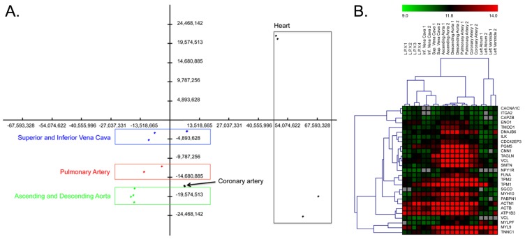
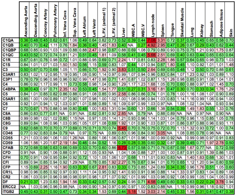
Genome-wide experiments are routinely used to increase the understanding of the biological processes involved in the development and maintenance of a variety of pathologies. Although the technical feasibility of this type of experiment has improved in recent years, data analysis remains challenging. In this context, gene set analysis has emerged as a fundamental tool for the interpretation of the results. Here, we review strategies used in the gene set approach, and using datasets for the pig cardiocirculatory system as a case study, we demonstrate how the use of a combination of these strategies can enhance the interpretation of results. Gene set analyses are able to distinguish vessels from the heart and arteries from veins in a manner that is consistent with the different cellular composition of smooth muscle cells. By integrating microRNA elements in the regulatory circuits identified, we find that vessel specificity is maintained through specific miRNAs, such as miR-133a and miR-143, which show anti-correlated expression with their mRNA targets.


Figures

Figure 1
Figure 1
Explicative scan portion of miRNA microarray after the RAKE and labeling reactions (A) and before hybridization (B). Spike-in spots are indicated by red lines; the blue arrow indicates a specific probe, and the orange arrow indicates its background probe. Each background probe was positioned to the right of its probe.
Figure 2
Figure 2
(A) Principal component analysis (PCA). The first three components account for 62.8% of the observed variance. The green rectangle identifies the group of ascending and descending aorta samples (green dots); the coronary artery is indicated by a black dot, the red rectangle highlights pulmonary artery samples (red dots), and the blue rectangle surrounds superior and inferior vena cava samples (blue dots). On the right, separated from other samples, are heart samples; (B) Heat map of muscle transcripts. Transcripts coding for muscle proteins are up-regulated in arteries with respect to veins. The red squares indicate up-regulated genes, and the green squares indicate down-regulated genes. The grey squares indicate genes for which no expression was detected. L.P.V. = leaflet of pulmonary valve; Inf. Vena Cava = inferior vena cava; Sup. Vena Cava = superior vena cava. The numbers following the sample names indicate the number of experimental replicates.
Figure 3
Figure 3
Expression of genes involved in the complement response. The numbers represent gene expression levels normalized to the average expression of the same gene across all tissues. Down-regulated genes are shown in green, and up-regulated genes are shown in red. Most of the up-regulated genes are expressed in the liver, which is responsible for the synthesis of most of the proteins of the complement system, in the spleen and in lymph nodes (lymphoid organs). NA = Expression not detected; L.P.V. = leaflet pulmonary valve; WBC.A = white blood cells from arterial blood; WBC.V = white blood cells from venous blood.
Figure 4
Figure 4
Regulatory network reconstructed using mutual information. The edges of the network are colored according to their prevalent expression. Heart-specific genes are shown in violet, vessel-specific genes are shown in blue, and genes without tissue-specific expression are shown in pink.
Figure 5
Figure 5
Gene and miRNA interaction sub-network describing vessel specificity. Triangles represent miRNAs; circles represent mRNAs. Gene expression in the ascending aorta according to log2 (gene expression/average gene expression) is represented by color; green indicates down-regulation, red indicates up-regulation. Under each node, histograms representing log2 (gene expression/average gene expression) in the ascending aorta, descending aorta, inferior vena cava, and superior vena cava (reading from left to right) are shown. The area highlighted by the circle indicates the densely connected portion of the sub-network (an enlarged view of this area is available in Figure 6).
Figure 6
Figure 6
Enlarged view of the densely connected area of Figure 5. (A) The colors indicate expression in the aorta; (B) The colors indicate expression in veins. The triangles represent miRNAs; circles represent mRNAs. Up-regulated = red; down-regulated = green; * = nodes discussed in the text; (C) qRT-PCR results confirm that there is an inverse relationship between miRNAs and their targets. P_15 is for prediction_15_14390446_14390503_-_3p. In Y axis the original expression level related to H3. Bars are for standard deviation between three replicates.
Figure 7
Figure 7
(A) Combination of pathway topology and ab initio reconstructed network. Nodes corresponding to the Reactome pathway (core nodes) are shown in red; additional genes in the first neighborhood of the core nodes obtained from the ab initio network are shown in light blue, and miRNAs are shown in grey; (B) Portion of (A) representing the miRNAs regulating the core nodes.

References

    1. Smyth G.K. Linear models and empirical bayes methods for assessing differential expression in microarray experiments. Stat. Appl. Genet. Mol. Biol. 2004;3:1–28. - PubMed
    1. Tusher V.G., Tibshirani R., Chu G. Significance analysis of microarrays applied to the ionizing radiation response. Proc. Natl. Acad. Sci. USA. 2001;98:5116–5121. - PMC - PubMed
    1. Shen K., Tseng G.C. Meta-analysis for pathway enrichment analysis when combining multiple genomic studies. Bioinformatics. 2010;26:1316–1323. - PMC - PubMed
    1. Callegaro A., Basso D., Bicciato S. A locally adaptive statistical procedure (LAP) to identify differentially expressed chromosomal regions. Bioinformatics. 2006;22:2658–2666. - PubMed
    1. Toedling J., Schmeier S., Heinig M., Georgi B., Roepcke S. MACAT—Microarray chromosome analysis tool. Bioinformatics. 2005;21:2112–2113. - PubMed

Publication types

LinkOut - more resources