. 2014 Jan 23;90(1):13.
doi: 10.1095/biolreprod.113.110197. Print 2014 Jan.

Nondividing, postpubertal rat sertoli cells resumed proliferation after transplantation

Affiliations

Nondividing, postpubertal rat sertoli cells resumed proliferation after transplantation

Payal Mital et al. Biol Reprod. .

Abstract

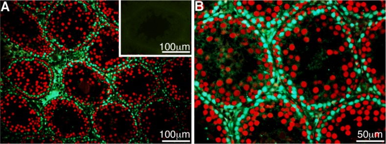
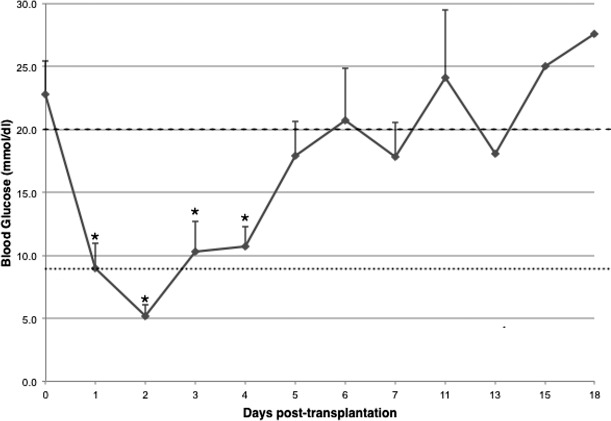
Conventionally, it was believed that Sertoli cells (SC) stopped proliferating at puberty and became terminally differentiated quiescent cells. However, recent studies have challenged that dogma. In this study, we transplanted nondividing SC isolated from 23- to 27-day-old postpubertal rats transduced with a recombinant adenoviral vector (containing furin-modified human proinsulin cDNA) into diabetic severe combined immunodeficiency mice. Immunostaining the grafts for cell proliferation markers, proliferating cell nuclear antigen (PCNA) and MKI67, revealed that transplanted SC within the grafts were proliferating. Possible causes for resumption of proliferation of SC could be viral transduction, cell isolation and culture, higher abdominal temperature at the transplant site, and/or transplantation. To test for these possible causes, double- immunofluorescence staining was performed for GATA4 (SC marker) and MKI67. None of the SC were positive for MKI67 in tissue collected during SC isolation and culture or at higher temperature. However, nontransduced SC stained positive for MKI67 after transplantation into rats, suggesting viral transduction was not a key factor for induction of SC proliferation. Interestingly, resumption in proliferative ability of nondividing SC was temporary, as SC stopped proliferating within 14 days of transplantation and did not proliferate thereafter. Quantification of 5-bromo-2'-deoxyuridine-labeled SC demonstrated that 7%-9% of the total transplanted SC were proliferating in the grafts. These data indicate for the first time that nondividing SC resumed proliferation after transplantation and further validate previous findings that SC are not terminally differentiated. Hence, transplantation of SC could provide a useful model with which to study the regulation of SC proliferation in vivo.

Keywords: Sertoli cells; proliferation; terminally differentiated; transplantation.


Figures

FIG. 1
FIG. 1
SC in 23- to 27-day-old Lewis rat testes were not proliferating. Testes were collected from 23- to 27-day-old Lewis rats, fixed in Z-Fix, and paraffin embedded. Tissue sections were immunostained for the SC marker GATA4 (green, A and B) and cell proliferation marker PCNA (red, A and B). B is the higher magnification of A. (A) Inset shows negative control for GATA4 and PCNA immunostaining.
FIG. 2
FIG. 2
Rat SC transduced with AdCMVhInsM caused a short-term lowering of BGL. SC were transduced with the recombinant adenoviral vector (at 100 MOI) containing furin-modified human proinsulin cDNA under the control of CMV promoter and transplanted into diabetic SCID mice. BGL were measured by obtaining blood from the tail vein of nonfasted SCID mice and the average BGL ± SEM values are shown. *Mean blood glucose values were significantly decreased compared to Day 0 (pretransplantation) values (P < 0.05). Dotted line represents normal BGL; dashed line represents the diabetic state.
FIG. 3
FIG. 3
Proliferation of transplanted SC is shown. SC cultured as monolayers were transduced with AdCMVhInsM (at 100 MOI). After 48 h, the transduced SC were collected and immunostained for insulin (A). Serial sections of graft-bearing kidneys collected from diabetic SCID mice were immunostained for insulin (B), the SC marker vimentin (C) or WT1 (E), and the cell proliferation marker PCNA (D) or MKI67 (F). Insets are higher magnifications (50 μm) of BD. B) Arrows indicate insulin positive SC. C and E) Arrows indicate vimentin- or WT1-positive SC that appear to be positive for PCNA or MKI67 (arrows, D and F). Hematoxylin was used as a counterstain to detect cell nuclei. A dotted line separates kidney cells from grafted cells.
FIG. 4
FIG. 4
Transplanted SC were positive for the proliferation markers PCNA and MKI67. Double immunofluorescence staining for GATA4 (green, A and D), PCNA (red, B) or MKI67 (red, E) was performed with tissue sections obtained from graft-bearing kidneys collected from diabetic SCID mice. C) Merged picture of GATA4 and PCNA is shown. F) Merged picture of GATA4 and MKI67 is shown. Arrows indicate cells that were double-positive for GATA4 and PCNA or MKI67. Insets show higher magnification of cells that were double-positive.
FIG. 5
FIG. 5
Cell isolation and culture did not induce SC proliferation. Double immunofluorescence staining was performed with tissue sections collected during cell isolation before collagenase treatment (AC) and after 2 days of culture (DF). SC were stained for GATA4 (green, A and D) and MKI67 (red, B and E). C and F) Merged pictures of GATA4 and MKI67 are shown.
FIG. 6
FIG. 6
Nondividing SC resumed proliferation after transplantation. SC were transplanted as syngeneic (AC) or allogeneic (DL) grafts. Graft-bearing kidneys were collected at Days 5 (AC), 10 (DF), 19 (G–I), and 55 (JL) post-transplantation, and tissue sections were double-immunostained for GATA4 (green, A, D, G, and J) and MKI67 (red, B, E, H, and K). C, F, I, and L) Merged pictures of GATA4 and MKI67 are shown. Arrows indicate cells that were double-positive for GATA4 and MKI67.
FIG. 7
FIG. 7
BrdU labeling of transplanted SC is shown. SC were transplanted in syngeneic rats (A and B) and immunocompromised mice (C and D). Tissue sections were double-immunostained for WT1 (green, AD) and BrdU (red, AD). B and D) Higher magnification of images shown in A and C, respectively. Arrows indicate cells that were double-positive for WT1 and BrdU.

Similar articles

Cited by

References

    1. Griswold MD. The central role of Sertoli cells in spermatogenesis. Semin Cell Dev Biol 1998; 9: 411–416. - PubMed
    1. Mital P, Kaur G, Dufour JM. Immunoprotective Sertoli cells: making allogeneic and xenogeneic transplantation feasible. Reproduction 2010; 139: 495–504. - PubMed
    1. Meinhardt A, Hedger MP. Immunological, paracrine and endocrine aspects of testicular immune privilege. Mol Cell Endocrinol 2011; 335: 60–68. - PubMed
    1. Chaudhary J, Sadler-Riggleman I, Ague JM, Skinner MK. The helix-loop-helix inhibitor of differentiation (ID) proteins induce post-mitotic terminally differentiated Sertoli cells to re-enter the cell cycle and proliferate. Biol Reprod 2005; 72: 1205–1217. - PubMed
    1. Orth JM. Proliferation of Sertoli cells in fetal and postnatal rats: a quantitative autoradiographic study. Anat Rec 1982; 203: 485–492. - PubMed

Publication types