. 2013 Nov 27;33(48):18987-98.
doi: 10.1523/JNEUROSCI.2394-13.2013.

Effects of reversible spinalization on individual spinal neurons

Affiliations

Effects of reversible spinalization on individual spinal neurons

Pavel V Zelenin et al. J Neurosci. .

Abstract

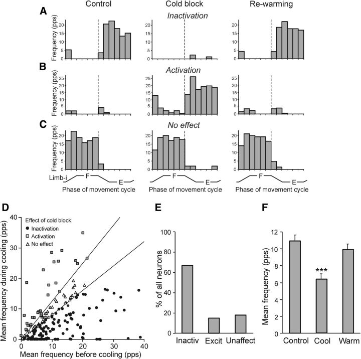
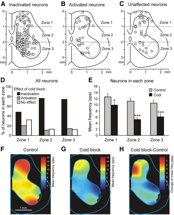
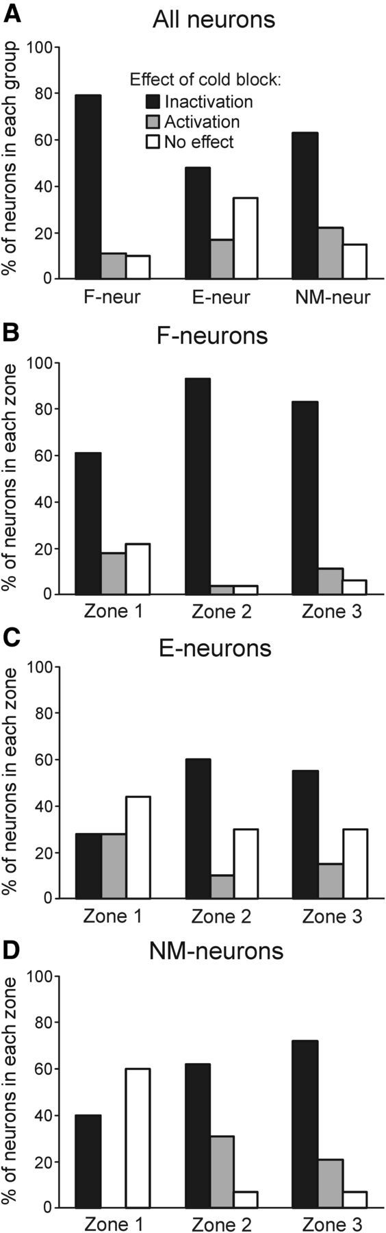
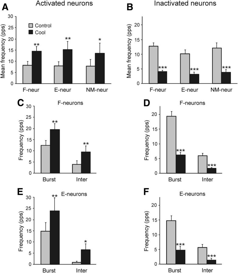
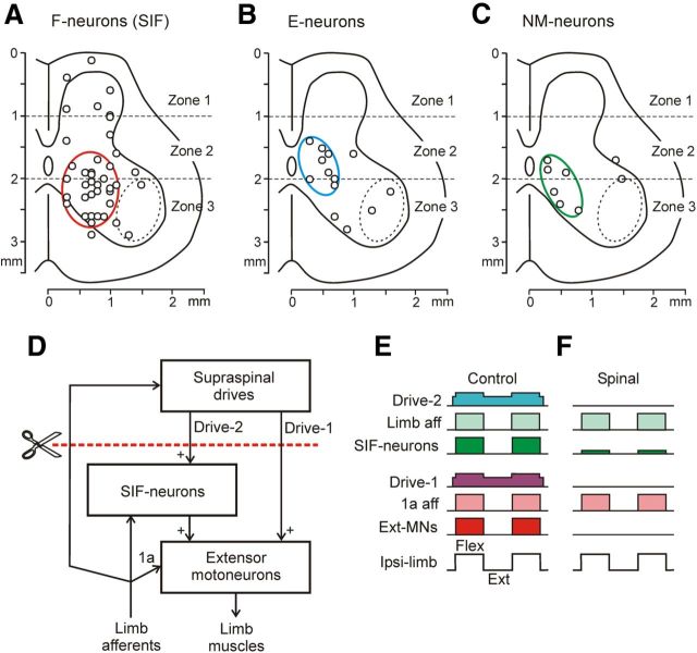
Postural limb reflexes (PLRs) represent a substantial component of the postural system responsible for stabilization of dorsal-side-up trunk orientation in quadrupeds. Spinalization causes spinal shock, that is a dramatic reduction of extensor tone and spinal reflexes, including PLRs. The goal of our study was to determine changes in activity of spinal interneurons, in particular those mediating PLRs, that is caused by spinalization. For this purpose, in decerebrate rabbits, activity of individual interneurons from L5 was recorded during stimulation causing PLRs under two conditions: (1) when neurons received supraspinal influences and (2) when these influences were temporarily abolished by a cold block of spike propagation in spinal pathways at T12 ("reversible spinalization"; RS). The effect of RS, that is a dramatic reduction of PLRs, was similar to the effect of surgical spinalization. In the examined population of interneurons (n = 199), activity of 84% of them correlated with PLRs, suggesting that they contribute to PLR generation. RS affected differently individual neurons: the mean frequency decreased in 67% of neurons, increased in 15%, and did not change in 18%. Neurons with different RS effects were differently distributed across the spinal cord: 80% of inactivated neurons were located in the intermediate area and ventral horn, whereas 50% of nonaffected neurons were located in the dorsal horn. We found a group of neurons that were coactivated with extensors during PLRs before RS and exhibited a dramatic (>80%) decrease in their activity during RS. We suggest that these neurons are responsible for reduction of extensor tone and postural reflexes during spinal shock.

Keywords: cold block; postural reflexes; rabbit; spinal cord injury; spinal neurons.


Figures

Figure 1.
Figure 1.
Experimental design. A, The decerebrate rabbit was fixed in a rigid frame (points of fixation are indicated by “X”). The cooler was positioned on the dorsal surface of the spinal cord at T12. To assess the conductance in spinal pathways under the cooler, the stimulating (Stim) and the recording (Rec) electrodes were inserted into the ventral spinal pathways at T11 and L1, respectively. Activity of spinal neurons from L5 was recorded by means of the microelectrode (ME). To evoke PLRs, the hindlimbs were positioned on a platform (B) periodically tilted in the transverse plane (C). The tilt of the platform caused flexion of one limb and extension of the other limb. The contact forces under the left and right limbs were measured by the force sensors (B, Force L and Force R, respectively). D, Limb configuration at the two extreme platform positions obtained from video recording (H, K, and An: hip, knee, and ankle joints, respectively). E, Time trajectory of the platform angle (Tilt) and of the vertical position of the distal point of the right and left limb (Limb-R and Limb-L, respectively).
Figure 2.
Figure 2.
Methods for reversible spinalization (A,B) and analysis of neuronal activity (C,D). A, Position of the cooler on the dorsal surface of the spinal cord, as well as positions of the stimulating (Stim) and recording (Rec) electrodes for monitoring the signal transmission in ventral spinal pathways. Black arrows indicate the flow of cooling agent through the cooler. B, Representative example of the effect of reversible spinalization on the signal transmission in ventral spinal pathways, on PLRs, and on the activity of a neuron recorded on the left side of L5. During this trial, periodical antiphase flexion/extension (F/E) movements of the left and right limbs were continuously performed by tilting the platform; the contact force and the EMG of vastus lateralis (Vast) were recorded bilaterally, along with the activity of the neuron. Trace Temp shows temperature of the cooler; arrowheads ON and OFF indicate the onset of cooling and the onset of rewarming, respectively. Insets Resp show response at L1 (marked by asterisk) to stimulation of T11 (100 μA, 0.1 ms pulse duration) performed at different time points (indicated by arrows 1–6). Note that disappearance of responses during cooling (point 3) was correlated with the disappearance of the force, EMG, and neuronal reactions to F/E limb movements, whereas reappearance of responses during rewarning (point 5) were correlated with restoration of these reactions. C, Raster of responses of the neuron shown in B in 10 sequential tilt cycles recorded before cooling. D, Histogram of spike activity of this neuron in different parts (1–12) of the tilt cycle. The halves of the cycle with higher and lower neuronal activity were designated as Burst and Interburst periods, respectively.
Figure 3.
Figure 3.
Effects of reversible spinalization on PLRs. A, Example of the left and right Vast and Force responses (elicited by antiphase F/E movements of both limbs). Responses were recorded before cooling (Control), during the cold block (Cool), and after rewarming (Warm). Under each condition, responses in 10 sequential tilt cycles were averaged. B, Mean values (±SE) of the dynamic and static force responses before cooling, during the cold block, after rewarming (n = 4, 16 tests, averaging over 80 responses for each condition), as well as the passive force (Psv, N = 4, n = 20). C, Example of the residual incorrectly phased response in Vast-L during the cold block. There was a 30× increase in EMG amplification during the cold block. D, Proportion of different types of EMG responses (elicited by antiphase F/E movements of both limbs) in Vast and Gast before cooling, during the cold block, and after rewarming. Flex, activation with ipsilimb flexion; Ext, activation with limb extensioin; Flex&Ext, activation with both movements; No resp, EMG not responding. Number of animals and tests: N = 10 and n = 51, respectively.
Figure 4.
Figure 4.
Population characteristics of three groups of spinal neurons (F-, E-, and NM-neurons) recorded before cooling. AC, Position of F-neurons (n = 109), E-neurons (n = 58), and NM-neurons (n = 32) on the cross-section of the spinal cord. An approximate position of motor nuclei is shown by the dotted line. Three zones of the gray matter are indicated: the dorsal (1), intermediate (2), and ventral (3) zones. DF, Average frequency in different phases of the movement cycle of the ipsilimb (mean ± SE) for F-neurons activated with limb flexion (D), for E-neurons activated with limb extension (E), and for NM-neurons not affected by platform tilts (F). G, Relative number of neurons with different coefficients of modulation. H, I, Relative number of F-neurons, E-neurons, and NM-neurons in all three zones of the gray matter together (H) and in each of the zones separately (I). The number of neurons recorded in zones 1–3: n = 56, n = 63, and n = 80, respectively.
Figure 5.
Figure 5.
Effects of reversible spinalization on individual spinal neurons. AC, Examples of the cold block effects: inactivation (A), activation (B), and no effect (C). D, Mean frequencies of individual neurons (n = 199) under two conditions: before cooling (abscissa) and during cold block (ordinate). The sector delineated the nonaffected neurons (in which the two frequencies differed by <20%). The neurons below the sector were considered inactivated; those above the sector activated. E, Proportion of neurons inactivated, activated, and unaffected by the cold block. F, Mean frequency of neurons before cooling (Control, n = 199), during the cold block (Cool, n = 199), and after rewarming (Warm, n = 187). ***p < 0.001.
Figure 6.
Figure 6.
Effect of reversible spinalization on the activity of different populations of spinal neurons. AC, Location of inactivated neurons (n = 134; A), activated neurons (n = 29; B), and unaffected neurons (n = 36; C). D, Proportion of inactivated, activated, and unaffected neurons in each of the three zones of the gray matter. E, Mean frequency of neurons in each of the three zones before cooling (Control) and during the cold block (Cool). *p < 0.05; ***p < 0.001. FH, Averaged distribution of neuronal mean frequencies on the cross-section of the spinal cord before cooling (F), during the cold block (G), and the difference between the two distributions (H). The frequency values are presented as a heat map (see Materials and Methods).
Figure 7.
Figure 7.
Effects of the reversible spinalization on different groups of spinal neurons. A, Proportion of F-, E-, and NM-neurons inactivated, activated, and unaffected by the cold block. BD, Proportion of F-neurons (B), E-neurons (C), and NM-neurons (D) inactivated, activated, and unaffected by cold block in each of three zones of the gray matter. The number of F-, E-, and NM-neurons recorded in zones 1–3: n = 33, 30, 46; n = 18, 20, 20; and n = 5, 13, 14, respectively.
Figure 8.
Figure 8.
Effects of the reversible spinalization on different characteristics of neuronal discharges. A, B, Effect on the mean frequency of activated (A) and inactivated (B) F-neurons, E-neurons, and NM-neurons. CF, Effect on the mean burst and interburst frequencies of F-neurons (C,D) and E-neurons (E,F) activated (C,E) and inactivated (D,F) by the cold block. *p < 0.05; **p < 0.01; ***p < 0.001.
Figure 9.
Figure 9.
A-C, Location of strongly inactivated neurons (>80% decrease of frequency) in different groups: F-neurons (A), E-neurons (B), and NM-neurons (C), on the cross section of the spinal cord. Areas with high density of these neurons are demarcated by red, blue, and green ellipses, respectively. DF, Presumed neuronal mechanisms underlying the disappearance of PLRs during spinal shock. D, Principal components of PLR mechanisms. E,F, Activity of these components in the nonspinalized (E) and spinalized (F) animals subjected to periodical platform tilts causing flexion/extension limb movements. SIF-neurons are the strongly inactivated F-neurons; the red interrupted line shows the level of spinalization. See text for explanations.

Similar articles

Cited by

References

    1. Apkarian AV, Hodge CJ, Jr, Martin RJ, Stevens RT. A cryogenic device for reversibly blocking transmission through small regions of the spinal cord white matter. J Neurosci Methods. 1989;29:93–106. doi: 10.1016/0165-0270(89)90020-4. - DOI - PubMed
    1. Ashby P, Verrier M. Neurophysiological changes following spinal cord lesions in man. Can J Neurol Sci. 1975;2:91–100. - PubMed
    1. Baldissera F, Hultborn H, Illert M. Integration in spinal neuronal systems. In: Brooks VB, editor. Handbook of physiology, Sec 1, The nervous system, Vol 2, Motor control. Bethesda, MD: American Physiological Society; 1981. pp. 509–595.
    1. Barnes CD, Joynt RJ, Schottelius BA. Motoneuron resting potentials in spinal shock. Am J Physiol. 1962;203:1113–1116. - PubMed
    1. Beloozerova IN, Zelenin PV, Popova LB, Orlovsky GN, Grillner S, Deliagina TG. Postural control in the rabbit maintaining balance on the tilting platform. J Neurophysiol. 2003;90:3783–3793. doi: 10.1152/jn.00590.2003. - DOI - PubMed

Publication types

LinkOut - more resources