. 2013 Nov 27;33(48):19012-22.
doi: 10.1523/JNEUROSCI.1797-13.2013.

Alleviating memory impairment through distraction

Affiliations

Alleviating memory impairment through distraction

Nathan Cashdollar et al. J Neurosci. .

Abstract

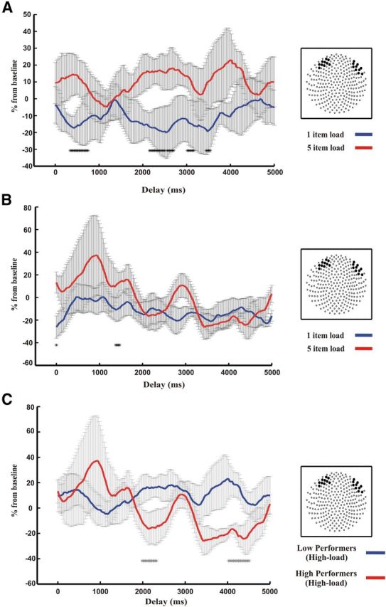
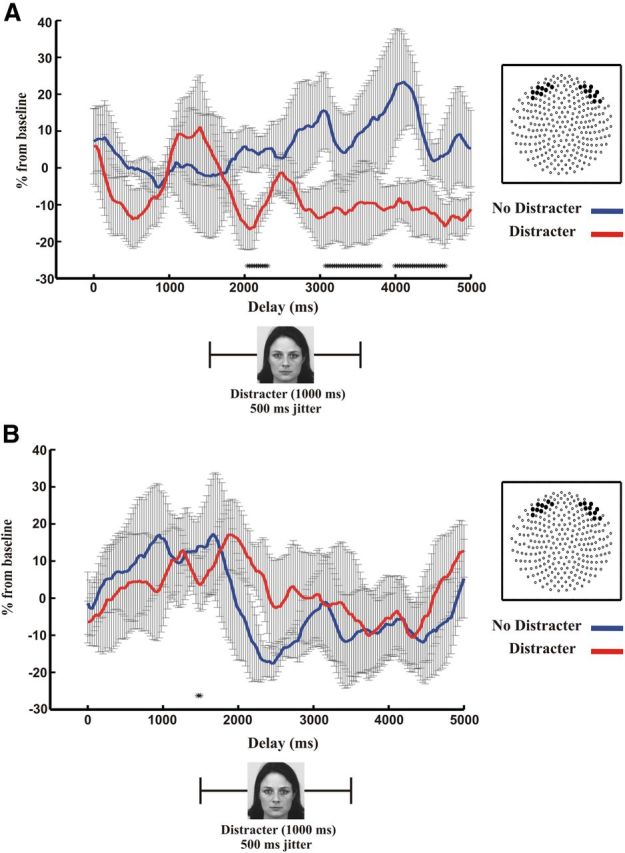
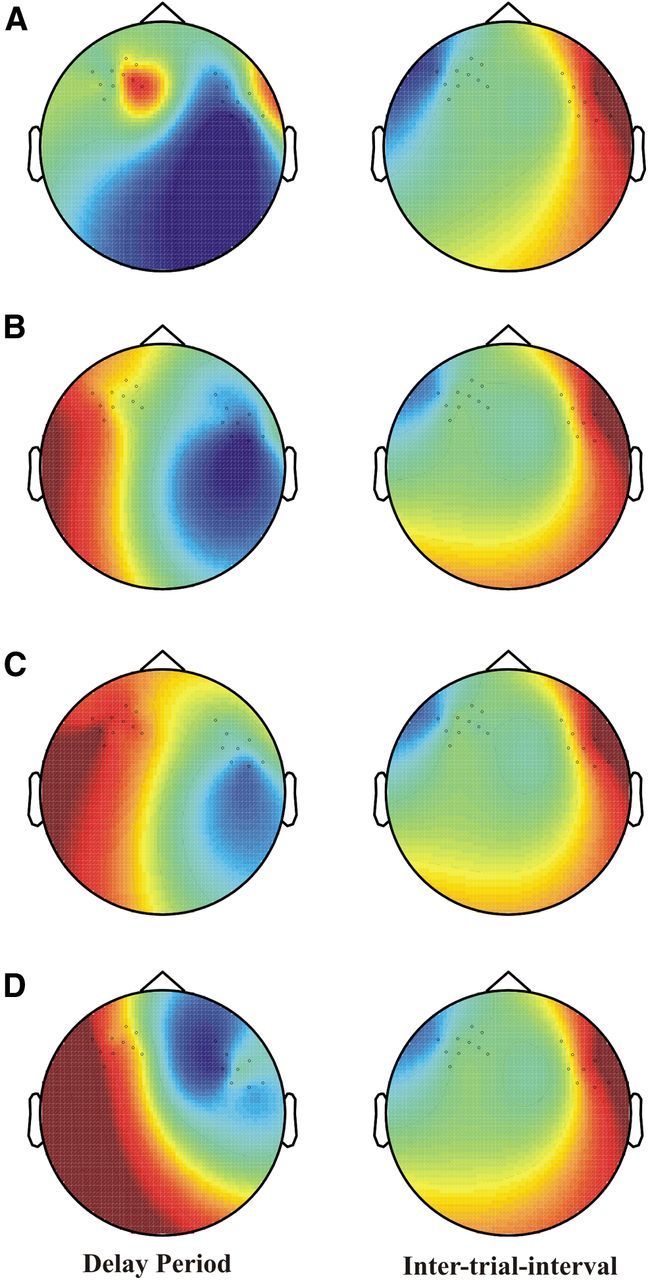
Distraction typically has a negative impact on memory for recent events and patients with existing memory impairment are particularly vulnerable to distractor interference. In contrast, here we establish a beneficial effect for distractor presentation in humans for both patients with memory impairment due to bilateral hippocampal lesions and healthy adults with low memory performance. Recognition memory for images of place scenes, which had to be memorized for short delay periods was significantly improved with the presentation of a distractor face during the delay. Magnetoencephalography recordings of neural oscillations in the theta frequency range obtained in healthy adults suggest that this memory improvement results from the interruption of rehearsal by the distractor. Our results highlight circumstances where active memory rehearsal may paradoxically increase memory impairments and distraction alleviates these memory deficits in patients with hippocampal injury and healthy adults.


Figures

Figure 1.
Figure 1.
Sample T1-weighted images of epilepsy cohorts. A, Epilepsy patient TE015 with isolated BHS and no other apparent structural or signal abnormalities. B, Patient FT026 with temporal lobe epilepsy determined to be MRI negative for hippocampal volume reductions and signal abnormalities (TLE).
Figure 2.
Figure 2.
Delayed match to sample (DMS) memory recognition tasks. Study 1 (top): An example of trials in which 1, 3, or 5 items are presented serially and must be maintained over a 5 s delay period in order to make a “match/no match” decision at test. Study 2 (bottom): An example of trials in which 5 items are presented serially and must be maintained over a 5, 20, or 45 s delay period to make a “match/no match” decision at test. In both Study 1 and Study 2, a face distractor was presented for 1 s with a temporal jitter in the middle of the delay on 50% of trials.
Figure 3.
Figure 3.
Delayed match to sample (DMS) memory load accuracy in Study 1. A, Comparisons of patients with BHS with NC1 normal control participants as a function of DMS memory load (1, 3, or 5 items) and distractor presence (D). When no distractor was present during the delay period (left), BHS patients' performance was significantly reduced compared with NC1; however, when a face distractor was presented for 1 s during the delay period (right), performance in the high-load conditions was enhanced such that differences to NC1 were attenuated to being nonsignificant. Comparable to the NC1 (younger and better educated) control group (B), BHS patients demonstrate similar memory load impairments for higher DMS load (3 and 5 items) conditions without distraction (left) compared with a group of age- and education-matched normal controls (NC2). These group differences are again alleviated to be nonsignificant when distraction was presented during the retention period (right) for the higher DMS load (3 and 5 items) conditions. C, Mean accuracy for DMS tasks for BHS patients (left) and patients with TLE but no hippocampal damage (right). When distraction was presented during the retention period (black), BHS patients' memory performance increased compared to when no distractor was presented (white). No such performance differences were found in the TLE group between distractor present and distractor absent conditions. Error bars indicate SEM. *p < 0.05; **p < 0.01; ***p < 0.001.
Figure 4.
Figure 4.
Study 2 investigated an additional group of normal controls (NC3) under high-load conditions (5 items) with extended memory delay durations (5, 20, or 45 s) to achieve similar levels of reduced memory performance as the BHS patients. A median split of the 45 s delay length condition without distraction (median value = 80%) resulted in two groups: a low performance group matched for accuracy to that of the BHS patients (A) and a high performance group (B). A, When the accuracy of the NC3 low performance group approached that of BHS patients for the high-load conditions (<80%), distractor presentation during the retention interval significantly improved memory performance. B, In the high performance group, no memory improvement was found with delay distraction. Error bars indicate SEM. **p < 0.01.
Figure 5.
Figure 5.
Bifrontal theta-band frequency synchrony in the high and low delayed match to sample (DMS) memory load conditions from Study 1. Serial-measures t test comparisons of 6 Hz phase coupling on selected sensor groups (right, insets) for low DMS memory load (1-item load in blue) versus high DMS memory load (5-item load in red) delayed memory recognition conditions of normal controls (NC1) in Study 1 (threshold of p < 0.05 per time point if present continuously over three successive theta cycles indicated by markings on x-axis). Error bars indicate SEM. A, In the low performers, bifrontal theta-coupling enhancement for the 5-item load condition continues throughout the memory delay compared with the 1-item load. However, in the high performers (B), bifrontal theta coupling is enhanced in the early portion of the delay period for 5-item load versus 1-item load and then diminishes over the course of the delay. C, Between-group comparisons of theta synchrony across a 5 s delay period of low performers (blue) versus high performers (red). Bifrontal theta coupling of the high performance group clearly declines over the course of the delay period, whereas in low performers, bifrontal theta coupling persists across the entire delay.
Figure 6.
Figure 6.
Bifrontal theta-band frequency synchrony of the high delayed match to sample (DMS) memory load (5 items) conditions with and without distraction for normal controls (NC1) in Study 1. Serial measures t test comparisons of 6 Hz phase coupling (threshold of p < 0.05 per time point if present continuously over three successive theta cycles indicated by markings on x-axis) on bifrontal sensor groups (right, insets). Error bars indicate SEM. A, Comparisons of theta synchrony across a 5 s delay period of low performers demonstrate that bifrontal theta coupling is reduced with distractor presentation (red) compared to without distractor presentation (blue). B, In comparison, distractor presence (red) in the high performance group show no differences between bifrontal theta coupling compared with the no distraction condition (blue) during the delay period.
Figure 7.
Figure 7.
Topographic distribution of event-related fields (ERFs) for the critical time window of analysis during the delayed match to sample (DMS) delay period (left) and the intertrial interval when participants were instructed to blink (right) on the same bifrontal sensors as in the primary phase-coupling analysis in NC1 (Study 1) for all relevant conditions of comparison (A, 1-item load in the low performers; B, 5-item load in the low performers; C, 5-item load with distraction in the low performers; and D, 5-item load in the high performers). The ERF topographies during the intertrial intervals clearly demonstrate the typical bifrontal distribution created by eye blink artifacts (right). In contrast, the critical DMS delay period time windows used for analysis (left) have a radically different distribution, demonstrating that ocular movements were not notably contaminating DMS delay period MEG recordings during any of the primary conditions of comparison used for the phase-coupling analysis.

Similar articles

Cited by

References

    1. Atkinson RC, Shiffrin RM. The control of short-term memory. Sci Am. 1971;225:82–90. doi: 10.1038/scientificamerican0871-82. - DOI - PubMed
    1. Axmacher N, Mormann F, Fernández G, Cohen MX, Elger CE, Fell J. Sustained neural activity patterns during working memory in the human medial temporal lobe. J Neurosci. 2007;27:7807–7816. doi: 10.1523/JNEUROSCI.0962-07.2007. - DOI - PMC - PubMed
    1. Baddeley A. Recent developments in working memory. Curr Opin Neurobiol. 1998;8:234–238. doi: 10.1016/S0959-4388(98)80145-1. - DOI - PubMed
    1. Baddeley A. Working memory and language: an overview. J Commun Disord. 2003;36:189–208. doi: 10.1016/S0021-9924(03)00019-4. - DOI - PubMed
    1. Cashdollar N, Malecki U, Rugg-Gunn FJ, Duncan JS, Lavie N, Düzel E. Hippocampal dependent and independent theta networks in human working memory. Proc Natl Acad Sci U S A. 2009;106:20493–20498. doi: 10.1073/pnas.0904823106. - DOI - PMC - PubMed

Publication types

MeSH terms

LinkOut - more resources