. 2013 Sep 16;15(5):R120.
doi: 10.1186/ar4300.

Propylthiouracil prevents cutaneous and pulmonary fibrosis in the reactive oxygen species murine model of systemic sclerosis

Propylthiouracil prevents cutaneous and pulmonary fibrosis in the reactive oxygen species murine model of systemic sclerosis

Gianluca Bagnato et al. Arthritis Res Ther. .

Erratum in

  • Arthritis Res Ther. 2014;16(2):406

Retraction in

Abstract

Introduction: Recent advances suggest that the cellular redox state may play a significant role in the progression of fibrosis in systemic sclerosis (SSc). Another, and as yet poorly accounted for, feature of SSc is its overlap with thyroid abnormalities. Previous reports demonstrate that hypothyroidism reduces oxidant stress. The aim of this study was therefore to evaluate the effect of propylthiouracil (PTU), and of the hypothyroidism induced by it, on the development of cutaneous and pulmonary fibrosis in the oxidant stress murine model of SSc.

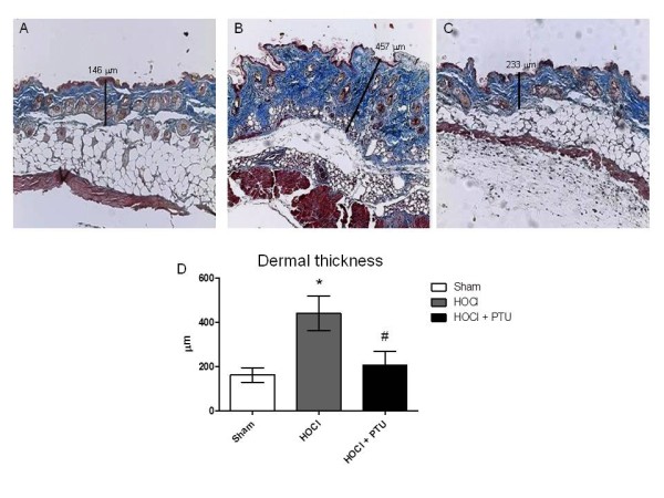
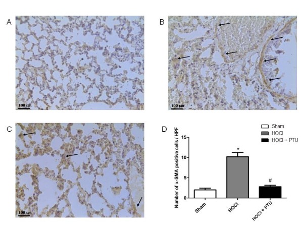
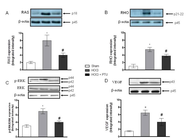
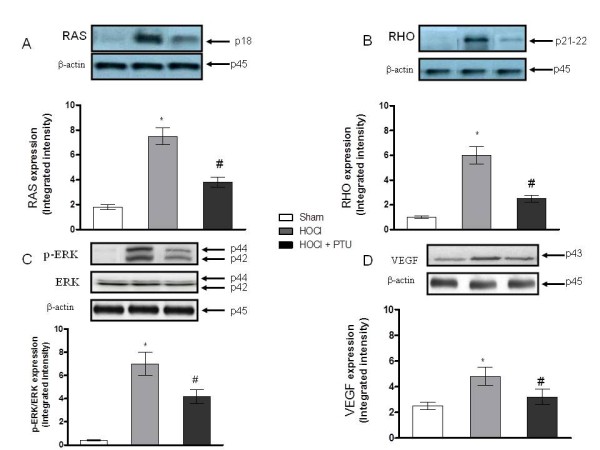
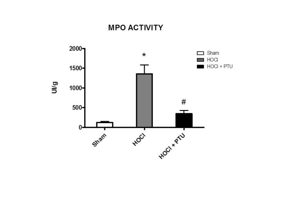
Methods: Chronic oxidant stress SSc was induced in BALB/c mice by daily subcutaneous injections of hypochlorous acid (HOCl) for 6 weeks. Mice (n = 25) were randomized into three arms: HOCl (n = 10), HOCl plus PTU (n = 10) or vehicle alone (n = 5). PTU administration was initiated 30 minutes after HOCl subcutaneous injection and continued daily for 6 weeks. Skin and lung fibrosis were evaluated by histologic methods. Immunohistochemical staining for alpha-smooth muscle actin (α-SMA) in cutaneous and pulmonary tissues was performed to evaluate myofibroblast differentiation. Lung and skin concentrations of vascular endothelial growth factor (VEGF), extracellular signal-related kinase (ERK), rat sarcoma protein (Ras), Ras homolog gene family (Rho), and transforming growth factor (TGF) β were analyzed by Western blot.

Results: Injections of HOCl induced cutaneous and lung fibrosis in BALB/c mice. PTU treatment prevented both dermal and pulmonary fibrosis. Myofibroblast differentiation was also inhibited by PTU in the skin and lung. The increase in cutaneous and pulmonary expression of VEGF, ERK, Ras, and Rho in mice treated with HOCl was significantly prevented in mice co-administered with PTU.

Conclusions: PTU, probably through its direct effect on reactive oxygen species or indirectly through thyroid function inhibition, prevents the development of cutaneous and pulmonary fibrosis by blocking the activation of the Ras-ERK pathway in the oxidant-stress animal model of SSc.


Figures

Figure 1
Figure 1
Accumulation of collagen in experimental dermal fibrosis is prevented by propylthiouracil administration. Dermal thickness was determined by using photomicrographs of Masson-stained sections, by measuring the distance between the epidermal-dermal junction and the dermal-fat junction at 10 randomly selected sites/high-power field (HPF), for 10 HPFs per section. Skin fibrosis was induced in mice by subcutaneous injection of HOCl. The resultant increase in dermal thickness was significantly reduced by subcutaneous injection of propylthiouracil. Representative Masson trichrome-stained sections were examined with light microscopy: (A) Normal histology of a representative skin tissue obtained from a Sham mouse; (B) Representative histology of skin tissue of HOCl mice; (C) Representative histology of skin tissue of HOCl + PTU mouse (original magnification,×10.); (D) Dermal thickness in mice from the three experimental groups (Sham group, n = 5; HOCl group, n = 10; HOCl + PTU group, n = 10). Values are expressed as the mean and SD. *P < 0.001 versus Sham #P < 0.001 versus HOCl.
Figure 2
Figure 2
Immunostaining for α-SMA (arrows, myofibroblasts nuclei) in cutaneous samples. Representative tissue sample from: (A) Sham animal; (B) HOCl mice; (C) HOCl + PTU animal (Original magnification, ×40). The arrows show strong diffuse staining of myofibroblasts nuclei (dark brown staining); (D) Number of myofibroblasts from the three experimental groups (HOCl + PTU group, n = 10; HOCl group, n = 10; Sham, n = 5). The increase of myofibroblast population in the skin of HOCl mice is prevented by propylthiouracil administration. Values are expressed as the mean and SD. *P < 0.001 versus Sham; #P < 0.001 versus HOCl.
Figure 3
Figure 3
Preventive effect of propylthiouracil administration upon pulmonary fibrosis development in HOCl-induced murine model of systemic sclerosis. Representative Masson's trichrome-stained section of lung examined by light microscopy: (A) Normal histology of a representative lung tissue from Sham mouse; (B) Representative lung section from HOCl mouse; (C) Representative lung section from HOCl + PTU mouse (Original magnification, ×10.); (D) Semiquantitative analysis of lung tissue graded by using the Ashcroft score, as described in Methods. The degree of pulmonary fibrosis was evaluated in Masson trichrome-stained sections by using the Ashcroft score (the grade of lung fibrosis was scored on a scale of 0 to 8 by using the following criteria: grade 0, normal lung; grade 1 to 2, minimal fibrous thickening of alveolar or bronchiolar wall; grade 3 to 4, moderate thickening of walls without obvious damage to lung architecture; grade 5 to 6, increased fibrosis with definite damage to lung structure; and grade 7 to 8, severe distortion of structure and large fibrous areas. Values are expressed as the mean and SD. *P < 0.001 versus Sham; #P < 0.001 versus HOCl. HOCl group (n = 10), HOCl + PTU group (n = 10), Sham (n = 5).
Figure 4
Figure 4
Immunostaining for α-SMA (arrows are illustrative for myofibroblasts nuclei) in pulmonary samples. Representative tissue sample from: (A) Sham animal; (B) HOCl mice; (C) HOCl + PTU animal (Original magnification, ×40). The arrows show strong diffuse staining of myofibroblasts nuclei (dark brown staining); (D) Number of myofibroblasts from the three experimental groups (HOCl + PTU group, n = 10; HOCl group, n = 10; Sham, n = 5). The increase of myofibroblast population in the skin of HOCl mice is prevented by propylthiouracil administration. Values are expressed as the mean and SD. *p < 0.001 versus Sham; #p < 0.001 versus HOCl.
Figure 5
Figure 5
Effect of propylthiouracil on RAS (A), RHO (B), pERK (C), and VEGF (D) proteins expression in lung tissue samples. Values in A through D are expressed by the mean and SD relative for each animal group. *P < 0.001 versus Sham; #P < 0.001 versus HOCl. HOCl group (n = 10), HOCl + PTU group (n = 10), Sham (n = 5).
Figure 6
Figure 6
Effect of propylthiouracil on RAS (A), RHO (B), pERK (C), VEGF (D) protein expressions in skin tissue samples. Values in A through D are expressed by the mean and SD relative for each animal group. *P < 0.001 versus Sham; #P < 0.001 versus HOCl. HOCl group (n = 10), HOCl + PTU group (n = 10), Sham (n = 5).
Figure 7
Figure 7
Myeloperoxidase (MPO) activation in the lungs is abrogated by propylthiouracil administration. MPO activity was defined as the quantity of enzyme degrading 1 μM hydrogen peroxide/minute at 37°C and was expressed in units per 100 mg of tissue. *P < 0.001 versus Sham; #P < 0.001 versus HOCl. HOCl group (n = 10), HOCl + PTU group (n = 10), Sham (n = 5).

References

    1. Wei J, Bhattacharyya S, Tourtellotte WG, Varga J. Fibrosis in systemic sclerosis: emerging concepts and implications for targeted therapy. Autoimmun Rev. 2011;15:267–275. doi: 10.1016/j.autrev.2010.09.015. - DOI - PMC - PubMed
    1. Gabrielli A, Svegliati S, Moroncini G, Pomponio G, Santillo M, Avvedimento EV. Oxidative stress and the pathogenesis of scleroderma: the Murrell's hypothesis revisited. Semin Immunopathol. 2008;15:329–337. doi: 10.1007/s00281-008-0125-4. - DOI - PubMed
    1. Dooley A, Shi-Wen X, Aden N, Tranah T, Desai N, Denton CP, Abraham DJ, Bruckdorfer R. Modulation of collagen type I, fibronectin and dermal fibroblast function and activity, in systemic sclerosis by the antioxidant epigallocatechin-3-gallate. Rheumatology (Oxford) 2010;15:2024–2036. doi: 10.1093/rheumatology/keq208. - DOI - PubMed
    1. Dooley A, Bruckdorfer KR, Abraham DJ. Modulation of fibrosis in systemic sclerosis by nitric oxide and antioxidants. Cardiol Res Pract. 2012;15:521958. - PMC - PubMed
    1. Tsou PS, Talia NN, Pinney AJ, Kendzicky A, Piera-Velazquez S, Jimenez SA, Seibold JR, Phillips K, Koch AE. Effect of oxidative stress on protein tyrosine phosphatase 1B in scleroderma dermal fibroblasts. Arthritis Rheum. 2012;15:1978–1989. doi: 10.1002/art.34336. - DOI - PMC - PubMed

Publication types

MeSH terms