. 2014 Aug;47(8):433-8.
doi: 10.5483/bmbrep.2014.47.8.153.

Stigmasterol isolated from marine microalgae Navicula incerta induces apoptosis in human hepatoma HepG2 cells

Affiliations

Stigmasterol isolated from marine microalgae Navicula incerta induces apoptosis in human hepatoma HepG2 cells

Young-Sang Kim et al. BMB Rep. 2014 Aug.

Abstract

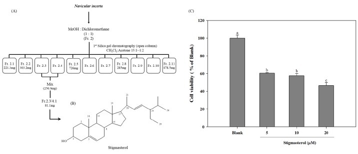
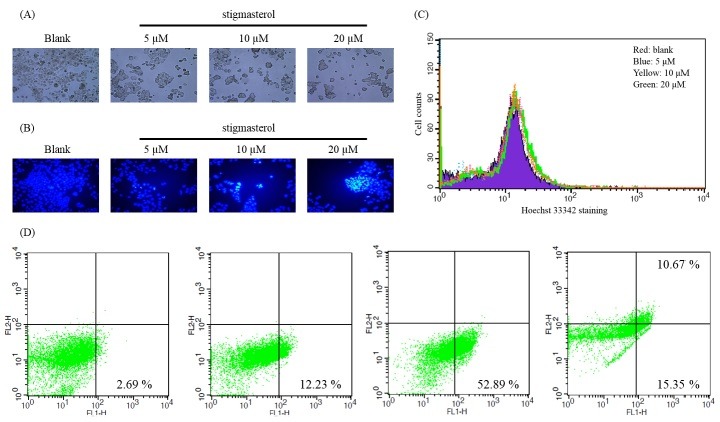
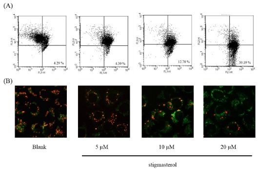
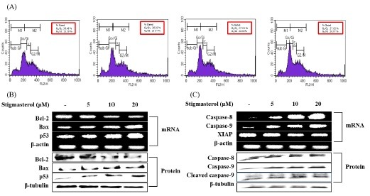
Plant sterols have shown potent anti-proliferative effects and apoptosis induction against breast and prostate cancers. However, the effect of sterols against hepatic cancer has not been investigated. In the present study, we assessed whether the stigmasterol isolated from Navicula incerta possesses apoptosis inductive effect in hepatocarcimona (HepG2) cells. According to the results, Stigmasterol has up-regulated the expression of pro-apoptotic gene expressions (Bax, p53) while down-regulating the anti-apoptotic genes (Bcl-2). Probably via mitochondrial apoptosis signaling pathway. With the induction of apoptosis caspase-8, 9 were activated. The DNA damage and increase in apoptotic cell numbers were observed through Hoechst staining, annexin V staining and cell cycle analysis. According to these results, we can suggest that the stigmasterol shows potent apoptosis inductive effects and has the potential to be tested as an anti-cancer therapeutic against liver cancer.


Figures

Fig. 1.
Fig. 1.. (A) Isolation profiles of stigmastsrol from Navicula incerta extract, (B) Structure of stigmasterol isolated from Navicula incerta. (C) Cell viability of HepG2 cells treated with stigmasterol for 24 h. Cells were cultured in serum-free media. After that HepG2 cells were treated with different concentrations (5, 10, 20 uM) of stigmasterol for 24 h and cell viability was assessed by the MTT assay. a-cDifferent letters on each bar indicates significant difference (P < 0.05) according to Duncan’s multiple range test.
Fig. 2.
Fig. 2.. (A) Morphological changes of stigmasterol treated HepG2 cells. For observation of morphological changes, cultured HepG2 cells were treated with stigmasterol for 24 h and morphological changes were detected under a light microscope (viewed at magnification of 100×). (B) Fluorescence micrographs showing the stigmasterol induced DNA damage. Stigmasterol treated cells were stained with Hoechst 33342 dye and detected under fluorescence microscope (viewed at magnification of 400×). The blue fluorescence in the nucleus indicates DNA fragmentation. (C) Cell numbers were counted using FACS after Hoechst 33342 staining. (Red; blank, Blue; 5 μM, Yellow; 10 μM, Green; 20 μM) (D) Flow cytometric analysis of the effect of stigmasterol in HepG2 cells experimented using Annexin V-PI staining assay.
Fig. 3.
Fig. 3.. Effect of stigmasterol on the mitochondrial membrane potential of HepG2 cells. (A) Fluorescence microscopic image of treated HepG2 cells stained with the MitoCaptureTM mitochondrial dye (Biovision). (B) Flow cytometric analysis of mitochondrial membrane potential.
Fig. 4.
Fig. 4.. Apoptotic inducing effect of stigmasterol in HepG2cells. Cells were treated with different concentrations of stigmasterol for 24 h. (A) Cell cycle progression patterns of HepG2 cells treated with stigmasterol. Stigmasterol treated cells were stained with PI and detected using FACS (FACS Calibur, BD Sciences, Heidelberg, Germany). (B) The expression levels of Bcl-2, Bax and p53, were detected using RT-PCR and western blot analysis, respectively. β-actin and β-tubulin were used as an internal standard. (C) The expression levels of caspase-8, -9 and XIAP were detected using RT-PCR and western blot analysis. β-actin and β-tubulin were used as an internal standard.

Similar articles

Cited by

References

    1. Spolaore P., Joannis-Cassan C., Duran E., Isambert A. Commercial applications of microalgae. J. Biosci. Bioeng. (2006);101:87–96. doi: 10.1263/jbb.101.87. - DOI - PubMed
    1. Plaza M., Herrero M., Cifuentes A., Ibanez E. Inovative natural functional ingredients from Microalgae. J. Agric. Food Chem. (2009);57:7159–7170. doi: 10.1021/jf901070g. - DOI - PubMed
    1. Kang K. H., Qian Z. J., Ryu B., Kim D., Kim S. K. Protective effects of protein hydrolysate from Marine Microalgae Navicula incerta on ethanol-induced toxicity in HepG2/CYP2E1 cells. Food Chem. (2011);132:677–685. doi: 10.1016/j.foodchem.2011.10.031. - DOI
    1. Ryan E., McCarthy F., Maguire A., O’Brien N. Phytosterol oxidation products: their formation, occurance and biological effects. Food Rev. Int. (2009);25:157–174. doi: 10.1080/87559120802682797. - DOI
    1. Awad A. B., Fink C. S. Phytosterols as anticancer dietary components: evidence and mechanism of action. J. Nutr. (2000);130:2127–2130. - PubMed

Publication types

MeSH terms