. 2013 Nov 25;27(4):462-8.
doi: 10.1016/j.devcel.2013.10.014.

Homotypic vacuole fusion in yeast requires organelle acidification and not the V-ATPase membrane domain

Affiliations

Homotypic vacuole fusion in yeast requires organelle acidification and not the V-ATPase membrane domain

Emily M Coonrod et al. Dev Cell. .

Abstract

Studies of homotypic vacuole-vacuole fusion in the yeast Saccharomyces cerevisiae have been instrumental in determining the cellular machinery required for eukaryotic membrane fusion and have implicated the vacuolar H(+)-ATPase (V-ATPase). The V-ATPase is a multisubunit, rotary proton pump whose precise role in homotypic fusion is controversial. Models formulated from in vitro studies suggest that it is the proteolipid proton-translocating pore of the V-ATPase that functions in fusion, with further studies in worms, flies, zebrafish, and mice appearing to support this model. We present two in vivo assays and use a mutant V-ATPase subunit to establish that it is the H(+)-translocation/vacuole acidification function, rather than the physical presence of the V-ATPase, that promotes homotypic vacuole fusion in yeast. Furthermore, we show that acidification of the yeast vacuole in the absence of the V-ATPase rescues vacuole-fusion defects. Our results clarify the in vivo requirements of acidification for membrane fusion.


Figures

Figure 1
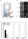
Figure 1. Acidification of the vacuole is required for vacuole morphology maintenance in vivo
(A) The V-ATPase is composed of a V1 ATP hydrolytic domain and a V0 membrane domain. The V0 domain is involved in H+ translocation and contains subunit a and the proteolipid ring composed of three different subunits in yeast. The highly conserved Arg at position 735 of subunit a is required for V-ATPase H+ translocation. (B) Parent (WT; KEBY136), vam3Δ (ECY157), vph1Δ (ECY145), and vph1R735Q (ECY149) cells expressing GFP-ALP (pLG230) as a vacuole maker were visualized by fluorescent (right column) or Differential Interference Contrast (DIC) microscopy (left column). Images shown are from a 0.5 μm Z-Stack. Scale bar = 5μm. (C) Quantification of the experiment shown in A. n=900 cells/strain, standard deviation is indicated. See also Figure S1.
Figure 2
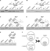
Figure 2. V0-dependent proton pumping is required on only one vacuole for homotypic fusion in vivo assessed using an ALP-processing assay
(A-E) Immunoblot analysis was used to assess whether ALP was present in its pro (pALP) or mature (mALP) form in lysates of diploids formed using the assay shown in F. Lysates of PEP4 (RPY10) and pep4-3 (SF838-9Da) haploid cells were included in the analysis to indicate the positions of mALP and pALP. In (A) and (B), the strains used are indicated. In (C), nyv1Δ strains (KEBY189 and KEBY192) were used and the presence or absence of the CUP1-NYV1 plasmid (pTC5) is indicated. Similarly, in (D) and (E), the strains used were vma3Δ (KEBY169 and KEBY171) and vph1Δ (ECY145 and ECY147), respectively, and the plasmids present were CUP1-VMA3 (pTC6) or CUP1-VPH1 (pGF669), as indicated. (F) Schematic representation of the ALP-processing assay to identify requirements of homotypic vacuole fusion in vivo. See also Figure S2.
Figure 3
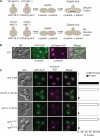
Figure 3. Vacuole acidification is required for fusion in vivo as assessed with a microscopic assay
(A) Schematic representation of a microscopic zygotic bud assay to identify requirements of homotypic vacuole fusion in vivo. In this schematic, fused vacuoles are depicted in white and unfused vacuoles are depicted in green or red. (B) Haploid parent cells expressing either GFPALP (pLG230) or mCherry-ALP (pMP2) were visualized by fluorescence or DIC microscopy during mating at the shmoo stage. Scale bar= 5μm (C) Haploid cells expressing either GFP-ALP or mCherry-ALP were mated in Yeast Extract Peptone with Dextrose (YEPD) and visualized by fluorescence and DIC microscopy. Large budded zygotes are shown for wild type x wild type (WT × WT, ECY153 and ECY155), vam3Δ x vam3Δ (ECY157 and ECY158), (vph1Δ x vph1Δ (ECY145 and ECY147), and vph1R735Q x vph1R735Q (ECY149 and ECY151). Buds are indicated by an asterisk, scale bar = 5Pm. Quantification of fusion in the zygotic bud in WT × WT (n=90), vam3Δ x vam3Δ (n=81), vph1Δ x vph1Δ (n=40), and vph1R735Q x vph1R735Q (n=51) are shown to the right of the cell images. Black bars indicate cells with fused vacuoles in the bud and white bars indicate zygotic buds with vacuoles that are not fused. Standard deviation is indicated.
Figure 4
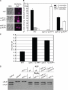
Figure 4. Expression of vacuole-localized Arabidopsis thaliana pyrophosphatase AVP1 rescues the vacuole fusion defect in cells lacking Vph1p
(A) WT (wild-type, SF838-1DD), vph1Δ + p:IPP1 (vph1::hyg ipp1 kanMX, ECY196 expressing plasmid-born IPP1 (p:IPP1, pEC139)), and vph1Δ + p:AVP1 (vph1::hyg ipp1 kanMX, ECY196 with plasmid pLG362 (p:AVP1) swapped for plasmid pEC139 (see supplemental methods)) and expressing mCherry-ALP (pGF242) were visualized using fluorescent and DIC microscopy. Scale bar = 5μm. (B) The vacuole morphology of WT (n=170), vph1Δ + p:IPP1 (ECY196, n=213), and vph1Δ cells + p:AVP1 (ECY196 with plasmid pLG362 swapped for plasmid pEC139, n=636) expressing plasmid borne mCherry-ALP (pGF242) was quantified. Standard deviation is indicated. (C) Vacuolar pH was measured in WT (n=4), vph1Δ + p:IPP1 (ECY196, n=5), and vph1Δ + p:AVP1 (ECY196 with plasmid pLG362 swapped for plasmid pEC139, n=5) using BCECF dye. Standard deviation is indicated. (D) Lysates of PEP4 (RPY10) and pep4-3 (SF838-9Da) haploid cells were included in the analysis to indicate the positions of mature ALP (mALP) and pro ALP (pALP) in the in vivo ALP content mixing assay. Processing of ALP is shown for vph1Δ diploids (ECY145 and ECY147), WT diploids (RYP10 and SF838-9Da), WT x vph1Δ diploids, vph1Δ ipp1Δ + p:AVP1 diploids (LGY253 and LGY254), and vph1Δ ipp1Δ + p:AVP1 x vph1Δ diploids. Plasmids present in ALP or Pep4p expressing cells in all lanes were pTC1 and pTC2.

References

    1. Baars TL, Petri S, Peters C, Mayer A. Role of the V-ATPase in regulation of the vacuolar fission-fusion equilibrium. Mol Biol Cell. 2007;18:3873–3882. - PMC - PubMed
    1. Baltscheffsky M, Schultz A, Baltscheffsky H. H+-proton-pumping inorganic pyrophosphatase: a tightly membrane-bound family. FEBS Lett. 1999;452:121–127. - PubMed
    1. Bayer MJ, Reese C, Buhler S, Peters C, Mayer A. Vacuole membrane fusion: V0 functions after trans-SNARE pairing and is coupled to the Ca2+-releasing channel. J Cell Biol. 2003;162:211–222. - PMC - PubMed
    1. Conradt B, Haas A, Wickner W. Determination of four biochemically distinct, sequential stages during vacuole inheritance in vitro. J Cell Biol. 1994;126:99–110. - PMC - PubMed
    1. Cowles CR, Snyder WB, Burd CG, Emr SD. Novel Golgi to vacuole delivery pathway in yeast: identification of a sorting determinant and required transport component. EMBO J. 1997;16:2769–2782. - PMC - PubMed

Publication types

MeSH terms