. 2013:4:2812.
doi: 10.1038/ncomms3812.

Cyclin D1 induction of Dicer governs microRNA processing and expression in breast cancer

Affiliations

Cyclin D1 induction of Dicer governs microRNA processing and expression in breast cancer

Zuoren Yu et al. Nat Commun. 2013.

Abstract

Cyclin D1 encodes the regulatory subunit of a holoenzyme that phosphorylates the pRB protein and promotes G1/S cell-cycle progression and oncogenesis. Dicer is a central regulator of miRNA maturation, encoding an enzyme that cleaves double-stranded RNA or stem-loop-stem RNA into 20-25 nucleotide long small RNA, governing sequence-specific gene silencing and heterochromatin methylation. The mechanism by which the cell cycle directly controls the non-coding genome is poorly understood. Here we show that cyclin D1(-/-) cells are defective in pre-miRNA processing which is restored by cyclin D1a rescue. Cyclin D1 induces Dicer expression in vitro and in vivo. Dicer is transcriptionally targeted by cyclin D1, via a cdk-independent mechanism. Cyclin D1 and Dicer expression significantly correlates in luminal A and basal-like subtypes of human breast cancer. Cyclin D1 and Dicer maintain heterochromatic histone modification (Tri-m-H3K9). Cyclin D1-mediated cellular proliferation and migration is Dicer-dependent. We conclude that cyclin D1 induction of Dicer coordinates microRNA biogenesis.


Conflict of interest statement

Competing Financial Interests: The authors declare no competing financial interests.

Figures

Figure 1
Figure 1. Endogenous cyclin D1 maintains Dicer expression
(A) Western blot showing decreased Dicer abundance in the cyclin D1 siRNA treated MCF-7 cells. Antibodies are directed to the miRNA biogenesis proteins Dicer, Ago2, DGCR8 and Drosha. (B) Real-time PCR analysis of Exportin 5 mRNA in MCF-7 cells treated with cyclin D1 siRNA or control siRNA. Values are equal to mean ±SEM (N=3). No significant difference between two samples by the standard two-tailed student’s t-test. (C) Real-time PCR demonstrating decreased cyclin D1 and Dicer mRNA level in the cyclin D1 siRNA treated MCF-7 cells. Values are equal to mean ±SEM of N=3 separate experiments.**p =0.0013 (cyclin D1) and 0.0026 (Dicer). Statistical significance was determined by the standard two-tailed student’s t-test. (D) Dicer mRNA abundance induced in the mammary gland of tet-inducable MMTV-cyclin D1a transgenic mice treated with doxycycline for 1 week. Data are mean ±SEM for N=3 separate transgenic mice *p=0.0334 (standard two-tailed student’s t-test). (E) Dicer protein level in mammary gland tumors of tet-inducible MMTV-cyclin D1a transgenic mice treated with doxycycline. (F) Dicer mRNA in mammary gland tumors of MMTV-cyclin D1a transgenic mice with comparison to age matched mammary gland. Data are mean ± SEM for 3 separate transgenic tumors and 3 normal mammary glands. *p=0.0308 (standard two-tailed student’s t-test). (G) Luciferase activity for the Dicer promoter in MCF-7 cells. Cells were treated with control and cyclin D1 siRNA as indicated. Data are mean ±SEM for triplicates in N=3 separate transfections normalized to β-galactosidase reporter activity. (H) Relative luciferase activity of the Dicer promoter and vector control in the presence of an expression vector for either cyclin D1 or a kinase-defective mutation (cyclin D1 KE). Data are shown as mean ±SEM for N=3 separate transfections. **p=0.0083 (cyclin D1a) and 0.0033 (cyclin D1KE). Statistical significance was determined by the standard two-tailed student’s t-test. (I) Cyclin D1 ChIP analysis revealed the specific binding to a region (−728 to −655) of the Dicer promoter. Both IgG IP to cyclin D1-overexpressing cells and cyclin D1-Flag IP to control cells were performed as negative control.
Figure 2
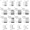
Figure 2. Dicer or cyclin D1 expression rescues the processing of pre-miRNA to mature miRNA in cyclin D1−/− cells
(A) Northern blot analysis of representative miRNAs (miR-16, let-7, miR-17, miR-20a) in WT and Dicer−/− HCT116 cells. (B–E) Northern blot analysis of pre-miR-16, pre-let-7a, pre-miR-17 and 20a to mature miRNA processing in cyclin D1+/+, cyclin D1−/− and cyclin D1−/−MEFs reintroduced with Dicer or cyclin D1a. (F–I) Northern blot detection of pre-let-7a, pre-miR-16, 17 and 20a and mature let-7a, miR-16, 17 and 20a in cyclin D1a (lane 1) and cyclin D1b (lane 2) rescued cyclin D1−/− MEFs (lane 3). (J) Quantitative analysis of pre-let-7a, pre-miR-16, 17 and 20a to mature let-7a, miR-16, 17 and 20a processing in cyclin D1−/− and cyclin D1+/+ MEFs. Values are equal to mean ± SEM (n=3). **p=0.0075 (miR-16), *p=0.0143 (let-7a), 0.05012 (miR-17) and 0.0472 (miR-20a). Statistical significance was determined by the standard two-tailed student’s t-test. tRNA serves as a loading control for northern blot.
Figure 3
Figure 3. Dicer and cyclin D1 govern heterochromatic H3K9 trimethylation
(A) HCT116 cells were stained with an antibody specific for trimethyl-H3K9 and DAP1 strained for nuclei. H3K9 trimethylation was assessed in Dicer+/+ vs. Dicer−/− HCT116 cells treated with cyclin D1 siRNA or control. Quantitative analysis of trimethyl-H3K9 induction by Dicer and cyclin D1 was indicated. Scale bars, 10 μm. mean ±SEM representing H3K9-Tri-me staining spots in 10 randomly selected cells. The experiments were repeated independently for three times. Statistical significance was determined by the standard two-tailed student’s t-test. (B) H3K9 trimethylation was assessed in cyclin D1+/+ vs. cyclin D1−/− MEFs. Scale bars, 10 μm. (C) Western blot for trimethyl-H3K9 in control and cyclin D1 siRNA treated MCF-7 cells. (D) Western blot for Dicer in WT and Dicer −/−HCT116 cells. (E) Western blot for cyclin D1 in WT and cyclin D1−/− MEFs. β-actin served as a loading control in all western blot analysis.
Figure 4
Figure 4. Dicer-dependent cyclin D1 induced cellular proliferation and migration
(A) Cellular proliferation was assessed using MTT assays on HCT116 Dicer+/+ and HCT116 Dicer−/− cells upon treatment with cyclin D1a siRNA or control siRNA as indicated. (B, C) Cellular migration assessed using wounding healing assays on Dicer−/− (B) or Dicer+/+ (C) HCT116 cells upon treatment with cyclin D1 siRNA or control siRNA as indicated. Scale bars 10 μm. (D) Quantitative analysis of the cellular migration assayed in B and C. A data are mean ±SEM representing three separate experiments with triplicate for each (N=9). B and C, Data are mean ±SEM for N=3 separate experiments.
Figure 5
Figure 5. A positive correlation analysis between Dicer and cyclin D1a expression in breast cancer patients
(A) Tree view display of gene expression from breast cancer patient samples for luminal A and basal genetic subtype. 2D-scatter plot for the elevated Dicer expression and high expression of cyclin D1a in luminal A breast cancer subtype (N=312). (B) 2D-scatter plot for the decreased Dicer expression and low expression of cyclin D1a in basal breast cancer subtype (N=359). The number of breast cancer patient samples found in each of the four quadrants is indicated. Plots are labeled with Pearson’s correlation coefficient R and the correlation p-value.
Figure 6
Figure 6. Hypothetical Mechanisms for cyclin D1 regulation of miRNA abundance
Schematic representation of the hypothetical molecular mechanisms by which cyclin D1a induces miRNA expression (A) or processing (B) through inducing Dicer expression and promoting pre-miRNA to mature miRNA processing. In turn, Dicer may involve in the regulation of cell proliferation and migration by cyclin D1 (C).

Similar articles

Cited by

References

    1. Bartel DP. MicroRNAs: genomics, biogenesis, mechanism, and function. Cell. 2004;116:281–297. - PubMed
    1. Yi R, Qin Y, Macara IG, Cullen BR. Exportin-5 mediates the nuclear export of pre-microRNAs and short hairpin RNAs. Genes & Development. 2003;17:3011–3016. - PMC - PubMed
    1. Yu Z, Wang C, Wang M, Li Z, Casimiro MC, Liu M, Wu K, Whittle J, Ju X, Hyslop T, et al. A cyclin D1/microRNA 17/20 regulatory feedback loop in control of breast cancer cell proliferation. The Journal of Cell Biology. 2008;182:509–517. - PMC - PubMed
    1. Lund E, Sheets MD, Imboden SB, Dahlberg JE. Limiting Ago protein restricts RNAi and microRNA biogenesis during early development in Xenopus laevis. Genes & Development. 2011;25:1121–1131. - PMC - PubMed
    1. Chen K, Rajewsky N. Deep conservation of microRNA-target relationships and 3′UTR motifs in vertebrates, flies, and nematodes. Cold Spring Harb Symp Quant Biol. 2006;71:149–156. - PubMed

Publication types

MeSH terms