. 2013 Nov 27;13(12):16216-33.
doi: 10.3390/s131216216.

Assessing the potential of low-cost 3D cameras for the rapid measurement of plant woody structure

Affiliations

Assessing the potential of low-cost 3D cameras for the rapid measurement of plant woody structure

Charles A Nock et al. Sensors (Basel). .

Abstract

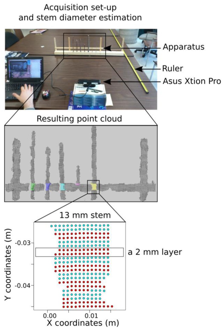
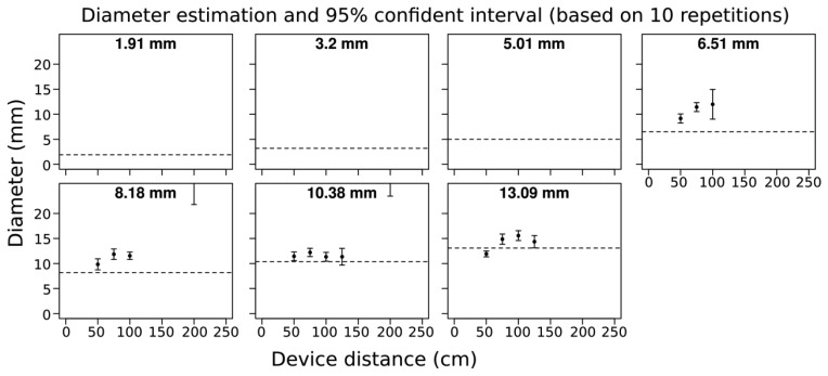
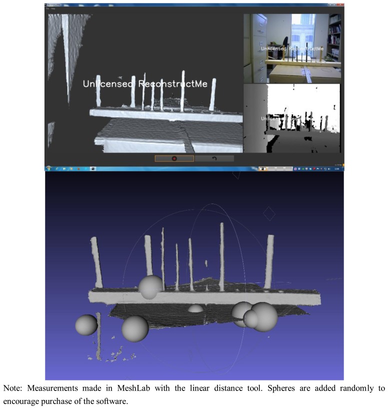
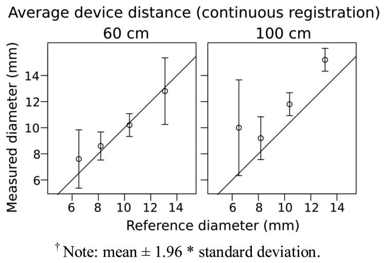
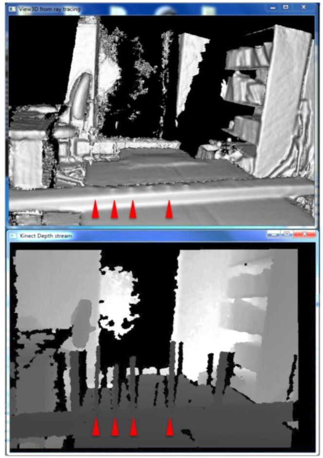
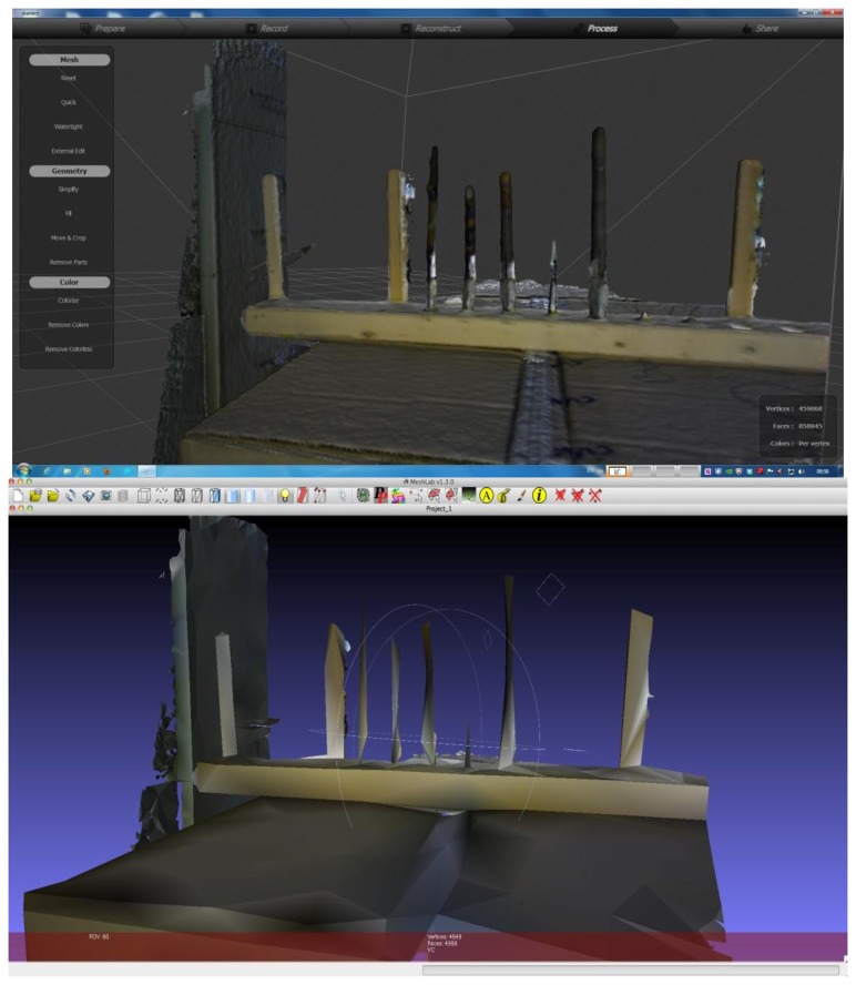
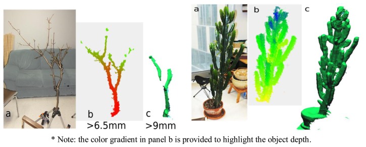
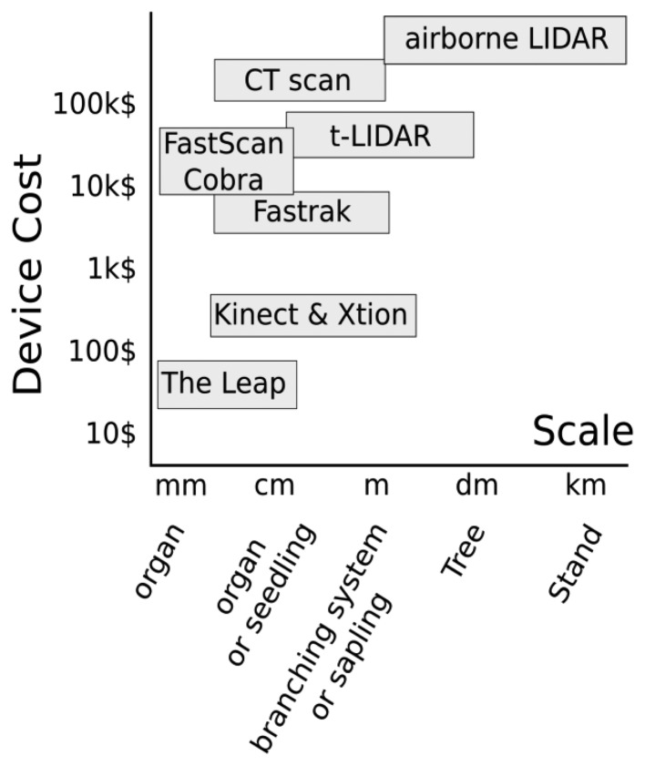
Detailed 3D plant architectural data have numerous applications in plant science, but many existing approaches for 3D data collection are time-consuming and/or require costly equipment. Recently, there has been rapid growth in the availability of low-cost, 3D cameras and related open source software applications. 3D cameras may provide measurements of key components of plant architecture such as stem diameters and lengths, however, few tests of 3D cameras for the measurement of plant architecture have been conducted. Here, we measured Salix branch segments ranging from 2-13 mm in diameter with an Asus Xtion camera to quantify the limits and accuracy of branch diameter measurement with a 3D camera. By scanning at a variety of distances we also quantified the effect of scanning distance. In addition, we also test the sensitivity of the program KinFu for continuous 3D object scanning and modeling as well as other similar software to accurately record stem diameters and capture plant form (<3 m in height). Given its ability to accurately capture the diameter of branches >6 mm, Asus Xtion may provide a novel method for the collection of 3D data on the branching architecture of woody plants. Improvements in camera measurement accuracy and available software are likely to further improve the utility of 3D cameras for plant sciences in the future.


Figures

Figure 1.
Figure 1.
Illustration of the experimental method for quantifying the threshold for diameter detection and the change in accuracy of diameter measurement with distance from static acquisitions with a 3D camera.
Figure 2.
Figure 2.
Threshold for diameter detection and accuracy of diameter measurement as a function of distance from branches of Salix of various diameters (shown in bold at center, top of each panel) measured from 3D images captured using the Asus Xtion Pro Live. Whiskers show 95% confidence intervals for the mean, calculated from 10 replicate acquisitions.
Figure 3.
Figure 3.
Top: screen capture of ReconstructMe showing the generated mesh (left), the RGB camera output (top right) and the depth camera output (bottom right). Bottom: resulting mesh from ReconstructME viewed in MeshLab. Spheres are added to generated meshes if a license has not been purchased. From left to right starting at the second vertical object: 7, 8, 10, 5, 13, 3 and 2 mm.
Figure 4.
Figure 4.
Comparison of reference and measured values for branches varying in diameter. Measured values were obtained from mesh generated using ReconstructMe and an Asus Xtion at 60 cm and 100 cm (other software tested did not yield measurable meshes). Circles show the mean of 5 scans and whiskers show 95% confidence intervals for the mean; 1:1 relationship between reference and measured values indicated with a solid line.
Figure 5.
Figure 5.
Example of depth stream (top) and resulting mesh (bottom) from the software KinFu and an Asus Xtion 3D camera. Notice that despite the presence of the branches in the depth stream that do not appear in the resulting mesh—suggesting that “filtering” of features of the generated mesh and their tenability will be important for continuous 3D plant scanning. Scanning time was <1 min.
Figure 6.
Figure 6.
Example of 3D scanning of woody branches using Skanect and an Asus Xtion. Top: original high-resolution mesh of woody branches scanned from ∼60 cm using Skanect. Bottom: a low-resolution mesh export is the only option available until a license is purchased.
Figure 7.
Figure 7.
Examples of 3D scanning of real plants. Maple branching system at left, and cactus-like euphorbia tree at right: (a) photograph; (b) point cloud based on 5 merged static scans *; (c) wire-frame meshes obtain by the continuous 3D scanning (KinFu software).
Figure 8.
Figure 8.
Illustration of devices used to generate 3D architectural data, their prices and the scale at which the generated data is most applicable to woody plants.

References

    1. Niinemets U., Valladares F. The Architecture of Plant Crowns: From Design Rules to Light Capture and Performance. In: Pugnaire F., Valladares F., editors. Functional Plant Ecology. Taylor and Francis; New York, NY, USA: 2007.
    1. Hopkinson C., Chasmer L., Young-Pow C., Treitz P. Assessing forest metrics with a ground-based scanning lidar. Can. J. For. Res. 2004;34:573–583.
    1. Lim K.S., Treitz P.M. Estimation of above ground forest biomass from airborne discrete return laser scanner data using canopy-based quantile estimators. Scand. J. For. Res. 2004;19:558–570.
    1. Yao T., Yang X., Zhao F., Wang Z., Zhang Q., Jupp D., Lovell J., Culvenor D., Newnham G., Ni-Meister W. Measuring forest structure and biomass in New England forest stands using Echidna ground-based lidar. Remote Sens. Environ. 2011;115:2965–2974.
    1. DeJong T.M., Da Silva D., Vos J., Escobar-Gutiérrez A.J. Using functional–structural plant models to study, understand and integrate plant development and ecophysiology. Ann. Bot. 2011;108:987–989. - PMC - PubMed

MeSH terms