Review
. 2014 May;406(14):3263-77.
doi: 10.1007/s00216-013-7473-1. Epub 2013 Nov 28.

Cellphone-based devices for bioanalytical sciences

Affiliations
Review

Cellphone-based devices for bioanalytical sciences

Sandeep Kumar Vashist et al. Anal Bioanal Chem. 2014 May.

Abstract

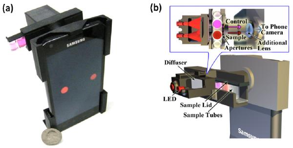
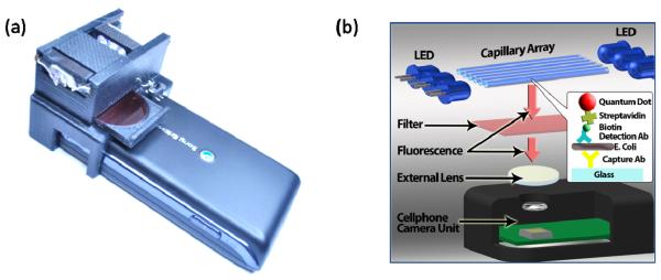
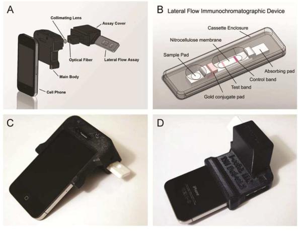
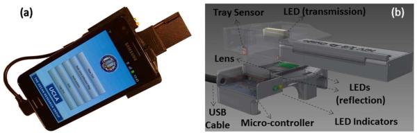
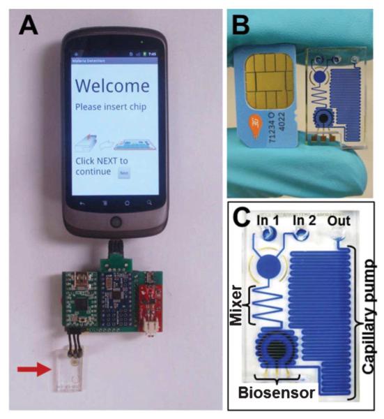
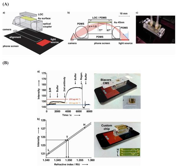
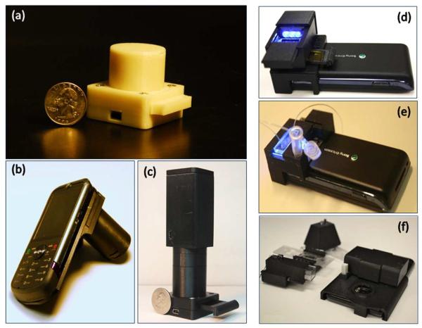
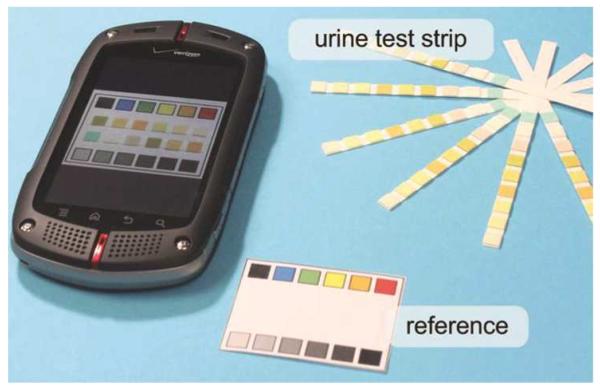
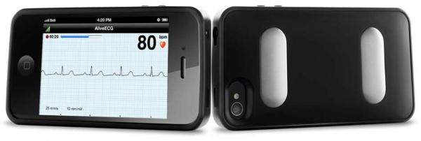
During the last decade, there has been a rapidly growing trend toward the use of cellphone-based devices (CBDs) in bioanalytical sciences. For example, they have been used for digital microscopy, cytometry, read-out of immunoassays and lateral flow tests, electrochemical and surface plasmon resonance based bio-sensing, colorimetric detection and healthcare monitoring, among others. Cellphone can be considered as one of the most prospective devices for the development of next-generation point-of-care (POC) diagnostics platforms, enabling mobile healthcare delivery and personalized medicine. With more than 6.5 billion cellphone subscribers worldwide and approximately 1.6 billion new devices being sold each year, cellphone technology is also creating new business and research opportunities. Many cellphone-based devices, such as those targeted for diabetic management, weight management, monitoring of blood pressure and pulse rate, have already become commercially-available in recent years. In addition to such monitoring platforms, several other CBDs are also being introduced, targeting e.g., microscopic imaging and sensing applications for medical diagnostics using novel computational algorithms and components already embedded on cellphones. This report aims to review these recent developments in CBDs for bioanalytical sciences along with some of the challenges involved and the future opportunities.


Figures

Fig. 1
Fig. 1
(A) Cellphone-based digital reader for colorimetric assays on iTube platform [5]. The opto-mechanical attachment (~22mm × 67mm × 75mm) that is installed at the back of the cellphone. (B) Schematics of the iTube platform. The iTube hardware attachment uses two interchangeable LEDs to illuminate the test and control tubes. Two diffusers are also inserted between the LEDs and the tubes to uniformly illuminate each tube. The transmitted light through each tube is then collected via a circular aperture and is imaged by the cellphone camera using an additional plano-convex lens (Focal length ~ 28 mm). A sample lid encloses the 6-well tube array and also works as an adaptor to insert the tube array inside the allergen tester attachment. Reproduced with permissions from the Royal Society of Chemistry.
Fig. 2
Fig. 2
(A) Cellphone-based E.coli detection platform. (B) Schematic diagram of the optical attachment for E. coli detection on a cellphone using a quantum dot-based sandwich assay embedded in glass capillary tubes [6]. Reproduced with permissions from the Royal Society of Chemistry.
Fig. 3
Fig. 3
(A) Enlarged view of the CBD showing placement of collimating lens and optical fiber set at specific angles in reference to the LFA cassette [8]. (B) LFA device utilizes a nitrocellulose membrane with gold conjugate and absorbing pads that are protected by a plastic cassette enclosure. Though capillary flow, the specimen (i.e., analyte) propagates to the other end of the membrane, labelled by the gold nano-particles in the gold conjugate pad. Test and control lines in the membrane are previously coated with antibodies specific to the target analyte, i.e., anti-TSH immobilized antibodies and anti-IgG immobilized antibodies, respectively. Forming the antibody-antigen complexes, the control and test lines develop a color change, which indicates the validity and the test results (positive/negative). (C,D) The actual reader attached to cellphone with an inserted disposable TSH LFA cassette. Reproduced with permission from Elsevier B.V.
Fig. 4
Fig. 4
(a) The universal Rapid Diagnostic Test (RDT) reader installed on an Android phone [9]. The light-weight (65 g) opto-mechanical attachment can be repeatedly attached/detached to the cellphone body without the need for fine alignment and modification. (b) Schematic of the designed optical RDT reader attachment. RDT tray works as a mechanical adaptor to insert various RDT types into the same cellphone based reader attachment. The tray sensor is a conductive component that is used to sense the insertion of the tray and ensures the proper operation of the device. Controlled using a simple micro-chip, the illumination LEDs uniformly illuminate the RDT of interest that is imaged by the cell-phone camera through an additional plano-convex lens. Reproduced with permission from the Royal Society of Chemistry.
Fig. 5
Fig. 5
(A) Assembled smartphone-based electrochemical sensor [15]. The arrow indicates the microfluidic chip. (B) Photograph of the chip and a mobile phone SIM card for comparison. (C) An enlarged image of the chip with labeled components. The channels are filled with a dye for improved visualization of the fluidic network. Reproduced with permission from the Royal Society of Chemistry.
Fig. 6
Fig. 6
(A) (a) 3D scheme of a representative setup for angle-resolved SPR using cellphone screen illumination and front camera detection optically coupled by a disposable device [21]. (b) 2D ray-trace of the experimental arrangement showing the light path from the screen to the camera. (c) Picture of the actual experimental arrangement. (B) (a) Interaction analysis of a commercial Biacore CM5 test chip functionalized for β2 microglobulin detection and tested at 1.32 μg mL−1 and 0.132 μg mL−1 concentrations. The baseline of the measurement is indicated with a blue line, while red and orange lines indicate normal serum and urine levels, respectively. (b) Test chip with embedded calibrations (H and L for high and low references) providing direct quantification of the unknown test value (T). Reproduced with permission from Wiley-VCH Verlag GmbH & Co. KGaA, Weinheim.
Fig. 7
Fig. 7
(a) A lensfree stand-alone microscope based on partially-coherent digital in-line holography [25]. (b) A lensfree cellphone microscope [26]. (c) Lensfree Holographic Pixel-Super Resolution Microscope [27]. (d) Picture of the fluorescent imager prototype utilizing an optical attachment for wide-field fluorescent imaging on a cellphone [28]. (e) Optofluidic fluorescent imaging cytometer on a cellphone [29]. (f) Cellphone based blood analysis platform. It includes a base attachment with two AA batteries and a universal port for adapting three different add-on components for white blood cell, red blood cell and hemoglobin density measurements [30]. Reproduced with permissions from The Royal Society of Chemistry and American Chemical Society.
Fig. 8
Fig. 8
Cellphone-based technique for colorimetric measurements in urine test strips [31]. Reproduced with permission from the Royal Society of Chemistry.
Fig. 9
Fig. 9
AliveCor's Heart Monitor [32]. It is an FDA approved device that has recently been cleared for sale in US for use by medical professionals to record, display, store, and evaluate single-channel electrocardiogram (ECG) rhythms. Reproduced with permission from David McCaman, AliveCor, Inc.

References

    1. [Accessed 4 July 2013]; http://www.itu.int/net/pressoffice/press_releases/2012/70.aspx#.UNl_qnfInjs.
    1. [Accessed 4 July 2013]; http://mobithinking.com/mobile-marketing-tools/latest-mobile-stats.
    1. McGeough CM, O'Driscoll S. IEEE Transactions on Biomedical Circuits and Systems. 2013. Camera Phone-Based Quantitative Analysis of C-Reactive Protein ELISA. DOI: 10.1109/TBCAS.2012.2234122. - PubMed
    1. Lu Y, Shi S, Qin J, Lin B. Low cost, portable detection of gold nanoparticle-labeled microfluidic immunoassay with camera cell phone. Electrophoresis. 2009;30:579–582. - PubMed
    1. Coskun AF, Wong J, Khodadadi D, Nagi R, Tey A, Ozcan A. A personalized food allergen testing platform on a cellphone. Lab Chip. 2013;13:636–640. - PMC - PubMed

Publication types