. 2013 Nov 28;4(11):e937.
doi: 10.1038/cddis.2013.454.

Differential regulation of the apoptotic machinery during megakaryocyte differentiation and platelet production by inhibitor of apoptosis protein Livin

Affiliations

Differential regulation of the apoptotic machinery during megakaryocyte differentiation and platelet production by inhibitor of apoptosis protein Livin

I Abd-Elrahman et al. Cell Death Dis. .

Abstract

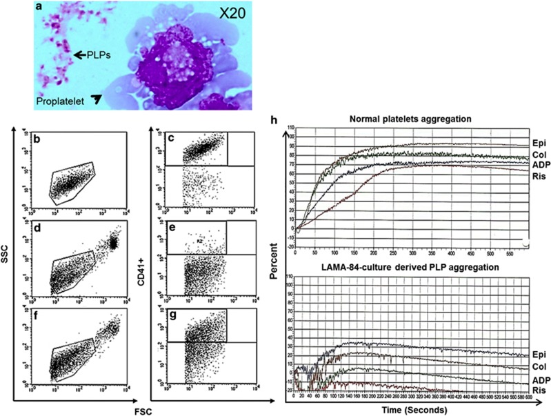
Livin is a member of the inhibitor of apoptosis proteins (IAP) family of intracellular antiapoptotic proteins that act by binding and inhibiting caspases. Upon strong apoptotic stimuli, it is then specifically cleaved by caspases to produce a truncated protein (tLivin) with a paradoxical proapoptotic activity. Intriguingly, we have detected robust protein levels of Livin in normal mature bone marrow megakaryocyte (MK) and platelets. To evaluate the potential role of Livin in thrombopoiesis, we used the human BCR-ABL+ cell line, LAMA-84, and cord blood CD34+ cells to induce differentiation toward MKs. Upon differentiation, induced by phorbol myristate acetate and concurrent with increase in Livin protein expression, LAMA-84 cells formed functional platelet-like particles. Livin overexpression in CD34+ progenitor cells induced higher endoreplication in the MKs generated. Furthermore, overexpression of Livin increased the ability of both primary MKs and differentiated LAMA-84 cells to produce functional platelets. In the differentiated LAMA-84 cells, we observed accumulation of proapoptotic tLivin concomitant with increased caspase-3 activity. Downregulation of Livin with small interfering RNA in both leukemic and primary MK cells decreased their ability to produce functional platelets. We suggest that Livin has a role in thrombopoiesis by regulating the apoptotic and antiapoptotic balance in MK endoreplication and platelet production.


Figures

Figure 1
Figure 1
Normal human MKs and platelets express Livin. (a) BM stained with secondary antibody and hematoxylin without Livin antibody as control. (b) Livin was detected in myeloid precursors and mature MKs in normal BM stained with a Livin-specific antibody. (c) Livin expression in MKs of a patient with idiopathic thrombocytopenic purpura (ITP) (original magnification, × 20). (d) The Livin protein was detected (by immunoprecipitation–western blot analysis) in platelets derived from the platelet concentrate (first two lanes). Both alternatively spliced variants of Livin, α and β, were overexpressed in LAMA-84 by transient transfection and used as a control (last two lanes) to compare endogenous Livin protein expression in platelets
Figure 2
Figure 2
Differentiation induction of LAMA-84 cells. LAMA-84 cells (0.5 × 10⁶/ml) were induced to differentiate to MKs by PMA (1.25–10 ng/ml) for 4 days. (a) May–Grunwald/Giemsa staining of cytospin preparations of cells without PMA treatment and following exposure to PMA (5 ng/ml). Note the proplatelet projections indicated by the arrow (original magnification, × 20). (b) Cells were stained with propidium iodide (50 μg/ml) and nuclear ploidy was measured by flow cytometry histograms from cultures without PMA and with PMA. (c and d) LAMA-84 cells (0.5 × 10⁶/ml) were cultured with PMA (5 or 10 ng/ml) to induce MK differentiation or with 50 μM Hemin to induce erythroid differentiation. Markers of MK differentiation, CD41 (MK marker) and CD71 (control early erythroid cell marker), were detected by flow cytometry (n=4). Undifferentiated (control) cells or Hemin-treated cells were of erythroid phenotype CD71+, whereas PMA-treated cells were primarily of the MK phenotype CD41+. (e) Livin, survivin and XIAP protein levels in the undifferentiated and differentiated LAMA-84 cells were evaluated by western blot. Note the appearance of Livin in PMA-treated cells (n=3)
Figure 3
Figure 3
Functional PLPs are released to the culture medium from differentiating cells. LAMA-84 cells (0.5 × 10⁶) were induced to differentiate to MKs by PMA (5 ng/ml). (a) On day 4, particles from cultured LAMA-84 cells were collected from media and stained with May–Grunwald/Giemsa. Both mature platelets and proplatelets are seen (original magnification, × 20). (b) Normal platelets from peripheral blood were used to establish the forward/side scatter (FSC/SSC) gates using flow cytometric. (c) Subsequent gating of CD41+ normal platelets. (d) Platelets of untreated LAMA84 cells were gated according their physical characteristic FSC/SSC set by control platelets and (e) Subsequent CD41 gating of PLPs from untreated LAMA-84 culture supernatants. (f) PLPs of PMA-treated LAMA-84 cells were gated by FSC/SSC for normal platelets and (g) subsequently by CD41 staining of PLPs from PMA-treated LAMA-84 culture supernatants. (h) Aggregation of normal platelets (top) and LAMA-84-culture derived PLPs (bottom) in response to epinephrine (Epi), collagen (Col), ADP and ristocetin
Figure 4
Figure 4
The proapoptotic activity of Livin is required for the generation of functional platelets. Control LAMA-84 cells (EV, empty vector) and LAMA-84 cells stably overexpressing WT Livin or Livin mutant lacking proapoptotic activity (Livin-RING) were treated with PMA for 4 days. Cells were harvested at 12, 24, 48 and 96 h. (a) Western blot analysis of Livin and XIAP proteins and their cleaved derivatives during LAMA-84 differentiation into MKs. Note the appearance of cleaved proapoptotic tLivin after 96 hours of differentiation induction with PMA. (b) Caspase-3 activation was assessed through the evaluation of caspase-3 enzyme activity (100 ng of protein) with Ac-DEVD-pNA (N-acetyl-Asp-Glu-Val-Asp-p-nitroaniline) in a colorimetric assay at day 4. Note that Livin transfection enhanced the caspase activity. (c) At day 4, culture-derived PLPs were collected from the medium for flow cytometric analysis of CD41 expression and the number of CD41+ platelet particles was quantitated. The PLP counts from cells transfected with either Livin or Livin-RING were significantly increased (n=3). (d) Functional assay for PLPs' aggregation in response to arachidonic acid (AA) was performed to determine the potential biological activity of these PLPs produced by LAMA-84 cells. Both control and Livin transfectants produced functional PLPs, whereas those derived from the Livin-RING-transfected cells were not functional (n=4)
Figure 5
Figure 5
Cord blood CD34+cells overexpressing Livin show increased differentiation toward the megakaryocytic lineage and higher ploidy. Cord blood CD34+ cells were cultured with TPO and SCF to drive MK differentiation. After one day, the cells were infected with control, WT Livin or RING mutant-expressing retroviruses. (a) At day 14, May–Grunwald/Giemsa staining of cytospin preparations showing polyploid MKs (n=6) (original magnification, × 60). (b) Dot plots of days 0 and 14 differentiated CD34+ cells stained for MK marker (CD41+, n=6). (c) CD41+ cell production was increased in CD34+ cells infected with Livin as compared with CD34+ cells incubated with TPO and SCF alone (n=6). (d) At day 14, the nuclei of MKs generated from CD34+ cells were counted in cytospins of cells stained with May–Grunwald/Giemsa to assess ploidy (n=6). (e) Western blot analysis of Livin protein expression in undifferentiated CD34+ cells (naive) and cells differentiated for 14 days with TPO and SCF and with infected empty vector or plasmid containing Livin. Note the appearance of cleaved proapoptotic tLivin after 14 days of differentiation
Figure 6
Figure 6
MKs derived from cord blood CD34+ cells overexpressing Livin or Livin-RING mutant produce more platelets in comparison with control. At day 14, platelets generated from MKs differentiated from CD34+ cells overexpressing either WT Livin, antiapoptotic-only Livin-RING or empty vector (control) were collected from the supernatant and analyzed for CD41 expression and platelet activation (n=6). (a) Platelets from cord blood were used to establish the gates for flow cytometric analysis. (b) Platelets generated from CD34+ cultures were similar in forward scatter, side scatter and CD41 expression. (c) The fold expansion of the percent of CD41+ platelets derived from CD34+ cells infected with WT Livin or antiapoptotic only Livin-RING as compared with empty vector. (d) Platelets derived from MK cultures (n=4) were incubated with ADP or arachidonic acid (AA) for 10 min at room temperature. Platelets were stained with anti-CD62p or isotype control to detect the activation of the platelets and analyzed on a flow cytometer. Iso, isotype control
Figure 7
Figure 7
The effect of the knockdown of Livin on platelets production and function. Downregulation of Livin expression was performed in both LAMA-84 cells (ac) and cord blood CD34+ cells (d and e) with the use of pSUPER-Livin-2 or control vector (pSUPER-Luc). (a) Western blot analysis showing the downregulation of both α- and β-Livin expression in LAMA-84 cells. (b) Knockdown of Livin in LAMA-84 cells reduced the number of PLPs (PLPs count/LAMA-84 cells count) (n=5). (c) Aggregation induced by various agonists (ADP (n=7), arachidonic acid (AA) (n=7), epinephrine (Epi) (n=4) and collagen (Col) (n=4)) is reduced in Livin-knockdown cells. (d) At day 14, the nuclei of MKs generated from CD34+ cells were counted in cytospins of cells stained with May–Grunwald/Giemsa (n=3). (e) Platelets derived from CD34+ cells differentiated toward MKs with and without the downregulation of Livin expression were incubated with ADP or AA and were stained with anti-CD62p to detected platelet activation (n=3). Iso, isotype control

References

    1. Gordge MP. Megakaryocyte apoptosis: sorting out the signals. Br J Pharmacol. 2005;145:271–273. - PMC - PubMed
    1. Ravid K, Lu J, Zimmet JM, Jones MR. Roads to polyploidy: The megakaryocyte example. J Cell Physiol. 2002;190:7–20. - PubMed
    1. Italiano JE, Lecine P, Shivdasani RA, Hartwig JH. Blood platelets are assembled principally at the ends of proplatelet processes produced by differentiated megakaryocytes. J Cell Biol. 1999;147:1299–1312. - PMC - PubMed
    1. Deutsch VR, Tomer A. Advances in megakaryocytopoiesis and thrombopoiesis: from bench to bedside. Br J Haematol. 2013;161:778–793. - PubMed
    1. Handagama PJ, Feldman BF, Jain NC, Farver TB, Kono CS. In vitro platelet release by rat megakaryocytes: effect of metabolic inhibitors and cytoskeletal disrupting agents. Am J Vet Res. 1987;48:1142–1146. - PubMed

Publication types

MeSH terms