. 2014 Jan;113(1):105-18.
doi: 10.1093/aob/mct262. Epub 2013 Nov 27.

Ontogenetic tissue modification in Malus fruit peduncles: the role of sclereids

Affiliations

Ontogenetic tissue modification in Malus fruit peduncles: the role of sclereids

Melanie Horbens et al. Ann Bot. 2014 Jan.

Abstract

Background and aims: Apple (Malus) fruit peduncles are highly modified stems with limited secondary growth because fruit ripening lasts only one season. They must reliably connect rather heavy fruits to the branch and cope with increasing fruit weight, which induces dynamic stresses under oscillating wind loads. This study focuses on tissue modification of these small, exposed structures during fruit development.

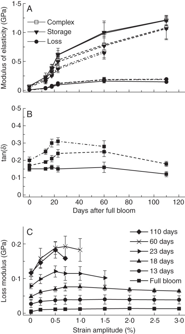
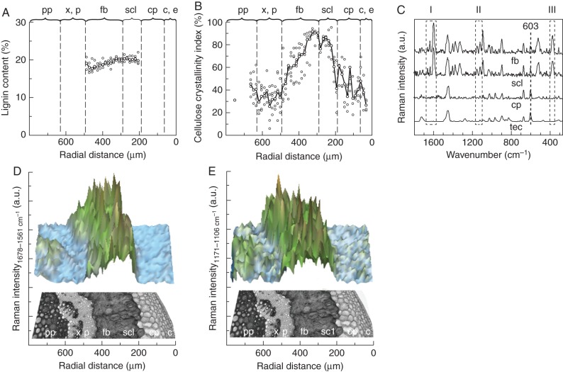
Methods: A combination of microscopic, static and dynamic mechanical tests, as well as Raman spectroscopy, was used to study structure-function relationships in peduncles of one cultivar and 12 wild species, representatively chosen from all sections of the genus Malus. Tissue differentiation and ontogenetic changes in mechanical properties of Malus peduncles were observed throughout one growing season and after successive removal of tissues.

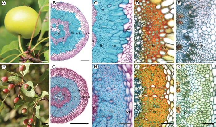
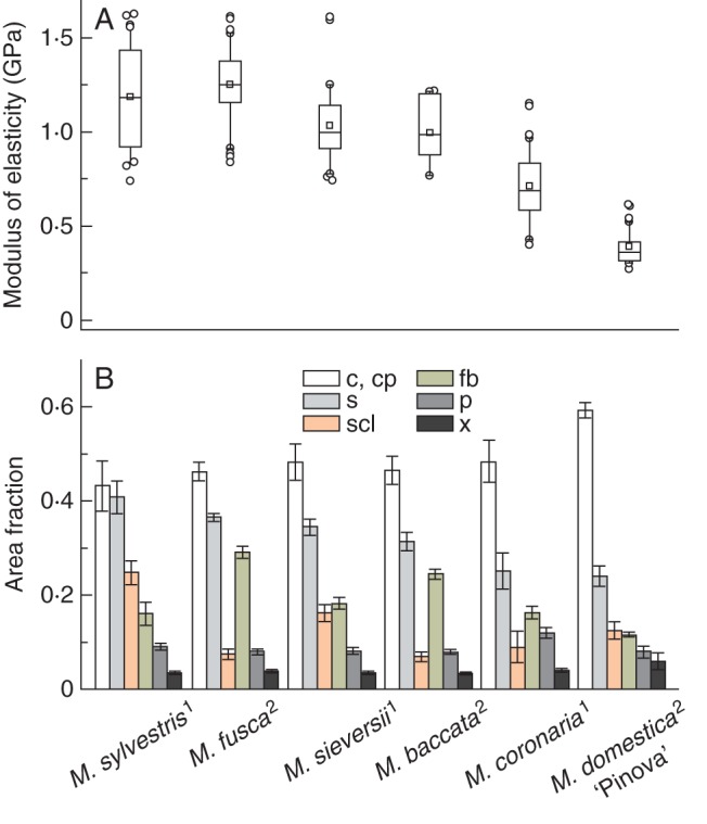
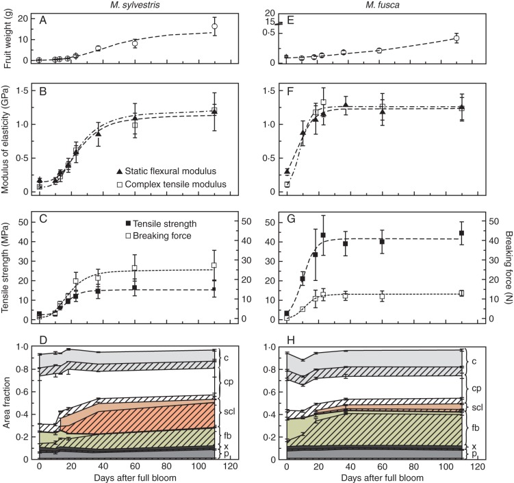
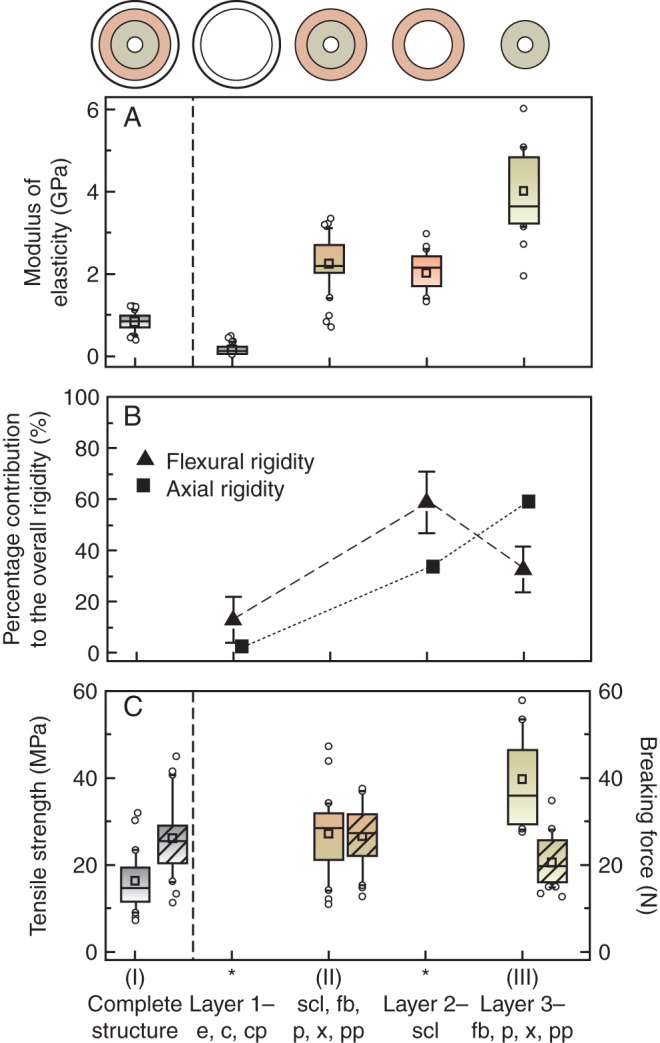
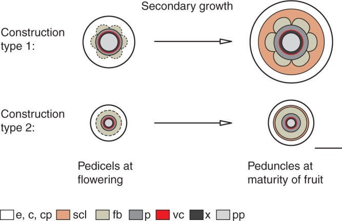
Key results: Unlike in regular stems, the vascular cambium produces mainly phloem during secondary growth. Hence, in addition to a reduced xylem, all species developed a centrally arranged sclerenchyma ring composed of fibres and brachysclereids. Based on differences in cell-wall thickness, and proportions and arrangement of sclereids, two types of peduncle construction could be distinguished. Fibres provide an increased maximum tensile strength and contribute most to the overall axial rigidity of the peduncles. Sclereids contribute insignificantly to peduncle strength; however, despite being shown to have a lower elastic modulus than fibres, they are the most effective tissue in stiffening peduncles against bending.

Conclusions: The experimental data revealed that sclereids originating from cortical parenchyma act as 'accessory' cells to enhance proportions of sclerenchyma during secondary growth in peduncles. The mechanism can be interpreted as an adaptation to continuously increasing fruit loads. Under oscillating longitudinal stresses, sclereids may be regarded as regulating elements between maintenance of stiffness and viscous damping, the latter property being attributed to the cortical parenchyma.

Keywords: Apple; Malus; biomechanics; fibres; fruit load; fruit peduncle; functional anatomy; sclereids; viscous damping.


Figures

Fig. 1.
Fig. 1.
Fruits and transverse sections of peduncles of M. sylvestris (construction type 1, A–E) and M. fusca (type 2, F–K) at different stages of fruit development. (B, C) Fibre caps in matured peduncles of type 1 are arranged in an open, flower-shaped ring – enclosed by a massive ring of thick-walled sclereids. (G, H) Mature peduncles of type 2 possess a closed ring of fibres – enclosed by a small layer of thin-walled sclereids. (D, J) Fibres with moderately thickened, lignified cell walls (primary cell-wall layers in green), and beginning formation of sclereids 18 d after full bloom. (E, K) In pedicels only scattered vessels are lignified. Staining: (B, C, G, H) Carmino Verte de Mirande; lignified tissues are stained green-blue, cellulose cell walls pink. (B, C, G, H) Wacker; lignified tissues are stained red–orange, cellulose cell walls green–blue. Scale bars: (A, F) = 10 mm; (B, G) = 200 µm; (C–E, H–K) = 50 µm. Abbreviations: c, collenchyma; cp, cortical parenchyma; cr, crystal clusters; e, epidermis; fb, fibres; p, phloem; pp, pith parenchyma; scl, sclereids; x, xylem.
Fig. 2.
Fig. 2.
(A) Structural modulus of elasticity obtained from static three-point bending tests of matured peduncles of different Malus accessions and types of construction according to Table 1 (index) ordered by magnitude. Central boxes represent the 25th and 75th percentiles with the median (line) and mean (square). Whiskers indicate the 10th and 90th percentile, and outliers are shown as circles. (B) Cross-sectional area fractions of single tissues of peduncles. High proportions of peripherally arranged cortical parenchyma with the highest contribution to the axial second moment result in decreased structural elastic modulus of the whole peduncle structure. Error bars are standard deviations. Abbreviations: c, collenchyma; cp, cortical parenchyma; fb, fibres; p, phloem; s, sclerenchyma (fibres and sclereids); scl, sclereids; x, xylem; 1, type 1; 2, type 2.
Fig. 3.
Fig. 3.
Comparison of peduncles of M. sylvestris (type 1, A–D) and M. fusca (type 2, E–H) during one growing season. Fruit weight, structural modulus of elasticity, maximum tensile strength and breaking force as well as cross-sectional area fractions of single tissues with their cell-wall area proportions are shown. The structural modulus of elasticity was calculated by applying strains of 0·05–0·25 % in static tests or 0·2 % in dynamic tests. Error bars are standard deviations; lines indicate the trends, based on non-linear fitting. The cross-sectional area fractions of single tissues are shown as different colours and their cell-wall area proportions are hatched. Abbreviations: c, collenchyma; cp, cortical parenchyma; e, epidermis; fb, fibres; p, phloem; scl, sclereids; x, xylem.
Fig. 4.
Fig. 4.
(A) Complex, storage and loss modulus, and (B) the loss factor of peduncles of M. sylvestris (type 1) during one growing season measured in dynamic tensile tests. Solid lines denote the values at strain amplitudes of 0·2 %, broken lines at 0·5 % and dashed/dotted lines at 1·0 %. (C) The loss modulus dependent on the strain amplitude and development stage (at a constant frequency of 1 Hz).
Fig. 5.
Fig. 5.
(A) Measured (I–III) and calculated (asterisks) modulus of elasticity, (B) proportional contribution of individual layers to overall flexural and axial rigidity (mean with SD) and (C) maximum tensile strength and breaking force (hatched) of the whole structure and single tissue layers of peduncles of M. sylvestris (type 1, from left to right). Central boxes represent the 25th and 75th percentiles with the median (line) and mean (squares). Whiskers indicate the 10th and 90th percentiles, and outliers are shown as circles. Abbreviations: c, collenchyma; cp, cortical parenchyma; e, epidermis; fb, fibres; p, phloem; pp, pith parenchyma; scl, sclereids; x, xylem.
Fig. 6.
Fig. 6.
(A) Distribution of lignin content and (B) the cellulose crystallinity index across a peduncle transverse section of M. sylvestris. (C) Representative Raman spectra of the embedding polymer Technovit and the peduncle tissues cortical parenchyma, sclereids and fibres normalized using the Technovit-specific peak at 603 cm−1 with marker bands for lignin (I), polysaccharides (II) and the cellulose crystallinity index (III). (D) Raman intensities calculated by integrating over defined wave number areas from 1678 to 1561 cm−1 for lignin and (E) from 1171 to 1106 cm−1 for polysaccharides. Abbreviations: c, collenchyma; cp, cortical parenchyma; e, epidermis; fb, fibres; p, phloem; pp, pith parenchyma; scl, thick-walled sclereids; tec, Technovit; x, xylem.
Fig. 7.
Fig. 7.
Schematic representation of the secondary growth of two different types of Malus peduncle construction. The increase in peduncle girth of type 1 due to cambial phloem formation entails cell elongations and division of cortical parenchyma by pushing outer tissues to the periphery. The fibre ring is displaced to the centre. The differentiation of cortical parenchyma cells outside the fibre ring into brachysclereids stiffens the structure most effectively against bending. Scale bar = 500 µm. Abbreviations: c, collenchyma; cp, cortical parenchyma; e, epidermis; fb, fibres; p, phloem; pp, pith parenchyma; scl, sclereids; vc, vascular cambium; x, xylem.

Similar articles

Cited by

References

    1. Agarwal UP, Reiner RS, Ralph SA. Cellulose I crystallinity determination using FT-Raman spectroscopy: univariate and multivariate methods. Cellulose. 2010;17:721–733.
    1. Altenbach H, Altenbach J, Rikards R. Einführung in die Mechanik der Laminat- und Sandwichtragwerke. 1st edn. Stuttgart: Deutscher Verlag für Grundstoffindustrie; 1996.
    1. Bogart SJ, Spiers G, Cholewa E. X-ray microCT imaging technique reveals corm microstructures of an arctic–boreal cotton-sedge, Eriophorum vaginatum. Journal of Structural Biology. 2010;171:361–371. - PubMed
    1. Boyd DW, Harris WM, Murry LE. Sclereid development in Camellia petioles. American Journal of Botany. 1982;69:339–347.
    1. Burgert I, Fratzl P. Plants control the properties and actuation of their organs through the orientation of cellulose fibrils in their cell walls. Integrative and Comparative Biology. 2009;49:69–79. - PubMed

Publication types