. 2012 Jan 5;5(1):79-93.
doi: 10.3390/ph5010079.

89Zr-Radiolabeled Trastuzumab Imaging in Orthotopic and Metastatic Breast Tumors

Affiliations

89Zr-Radiolabeled Trastuzumab Imaging in Orthotopic and Metastatic Breast Tumors

Albert J Chang et al. Pharmaceuticals (Basel). .

Abstract

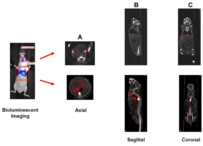
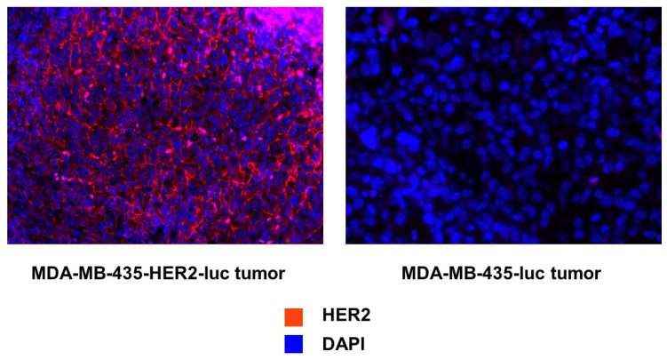
The human epidermal growth factor receptor 2 (HER2/neu) is overexpressed in 20-30% of breast cancers and is associated with tumor growth, angiogenesis, and development of distant metastases. Trastuzumab, an anti-HER2 monoclonal antibody, is used for the treatment of HER2 positive breast cancer and clinical efficacy of this agent is dependent on HER2 expression. Targeted PET imaging of HER2 with radiolabeled trastuzumab may be used to determine HER2 expression levels and guide therapy selection. The purpose of the current study was to evaluate a facile 89Zr-trastuzumab preparation method that can be efficiently applied for clinical grade production. Also, relative HER2 expression levels in orthotopic and metastatic breast cancer models were assessed by PET imaging using the 89Zr-trastuzumab produced by this simpler method.


Figures

Figure 1
Figure 1
Flow cytometric analysis of HER2/neu expression. The MDA-MB-435-HER2-luc and MDA-MB-435 breast cancer cell lines were evaluated for HER2/neu expression. Trastuzumab was used as the primary antibody, and FITC-conjugated goat anti-human IgG was used as the secondary antibody. Data are shown as cell number on the ordinate access and HER2/neu intensity on the abscissa.
Figure 2
Figure 2
.Cell binding studies. (A) Plot of the (total/bound) activity versus (1/[normalized cell concentration]), used to calculate the immunoreactive fraction of 89Zr-Df-NCS-trastuzumab in MDA-MB-435-Her2-luc cells by extrapolation to the y-intercept; (B) Cell uptake curves of Fraction bound versus normalized cell concentration in MDA-MB-435-Her2-luc and MDA-MB-435-luc. A blocking dose of unlabeled trastuzumab inhibited the binding of 89Zr-radiolabeled trastuzumab.
Figure 3
Figure 3
Biodistribution of 89Zr-radiolabeled trastuzumab in MDA-MB-435-Her2-luc and MDA-MB-435-luc orthotopic xenograft models at (A) 24 h and (B) 96 h post-injection. Data are expressed as percent injected dose per gram ± standard deviation, n = 5 for each time point.
Figure 4
Figure 4
Representative microPET/CT images obtained at 24 and 96 h in (A) and (B) respectively. Tumors implanted in the right mammary fat pad of each mouse. The axial (1), sagittal (2), and coronal (3) slices at the center of the tumor are demonstrated along with the maximal intensity projection reconstruction (4). The scale expressed as standardized uptake value (SUV) is demonstrated at the far right.
Figure 5
Figure 5
Detection of Her2+ metastatic lesions. 2 × 10⁶ MDA-MB-435-Her2-luc cells were administered via tail vein injection. Six weeks later, bioluminescent imaging (far right panel) demonstrated development of metastasis in the head and thoracic region. MicroPET/CT imaging using 89Zr-radiolabeled trastuzumab was able to localize these lesions to the L jaw and thoracic vetebral body. Shown are the axial (A), sagittal (B), and coronal (C) slices at the center of the tumor.
Figure 6
Figure 6
Immunofluorescent staining to evaluate the relative levels of HER2/neu expression for each tumor type. HER2/neu staining is demonstrated in red with a counterstain for the nucleus in blue.

References

    1. Citri A., Yarden Y. EGF-ERBB signalling: Towards the systems level. Nat. Rev. Mol. Cell Biol. 2006;7:505–516. doi: 10.1038/nrm1962. - DOI - PubMed
    1. Slamon D.J., Clark G.M., Wong S.G., Levin W.J., Ullrich A., McGuire W.L. Human breast cancer: Correlation of relapse and survival with amplification of the HER-2/neu oncogene. Science. 1987;235:177–182. - PubMed
    1. Slamon D.J., Godolphin W., Jones L.A., Holt J.A., Wong S.G., Keith D.E., Levin W.J., Stuart S.G., Udove J., Ullrich A., et al. Studies of the HER-2/neu proto-oncogene in human breast and ovarian cancer. Science. 1989;244:707–712. - PubMed
    1. Ignatiadis M., Desmedt C., Sotiriou C., de Azambuja E., Piccart M. HER-2 as a target for breast cancer therapy. Clin. Cancer Res. 2009;15:1848–1852. doi: 10.1158/1078-0432.CCR-08-1844. - DOI - PubMed
    1. Whenham N., D'Hondt V., Piccart M.J. HER2-positive breast cancer: From trastuzumab to innovatory anti-HER2 strategies. Clin. Breast Cancer. 2008;8:38–49. doi: 10.3816/CBC.2008.n.002. - DOI - PubMed