. 2014 Feb;65(2):527-38.
doi: 10.1093/jxb/ert396. Epub 2013 Nov 28.

Dual regulation of cytosolic ascorbate peroxidase (APX) by tyrosine nitration and S-nitrosylation

Affiliations

Dual regulation of cytosolic ascorbate peroxidase (APX) by tyrosine nitration and S-nitrosylation

Juan C Begara-Morales et al. J Exp Bot. 2014 Feb.

Abstract

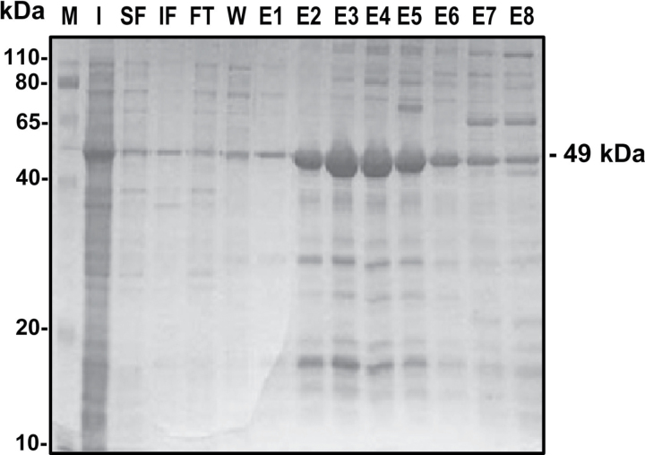
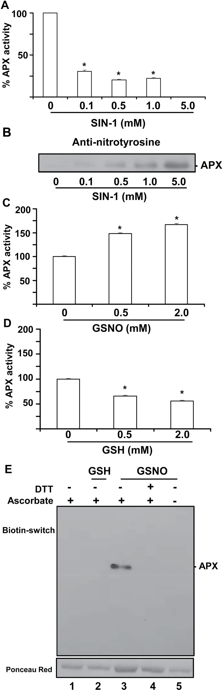
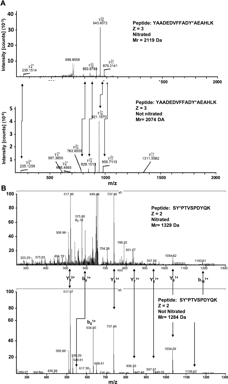
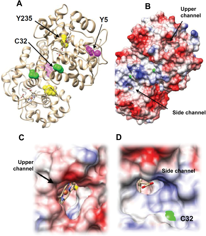
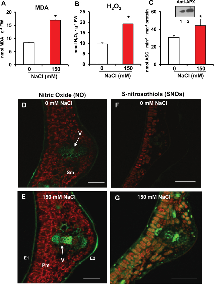
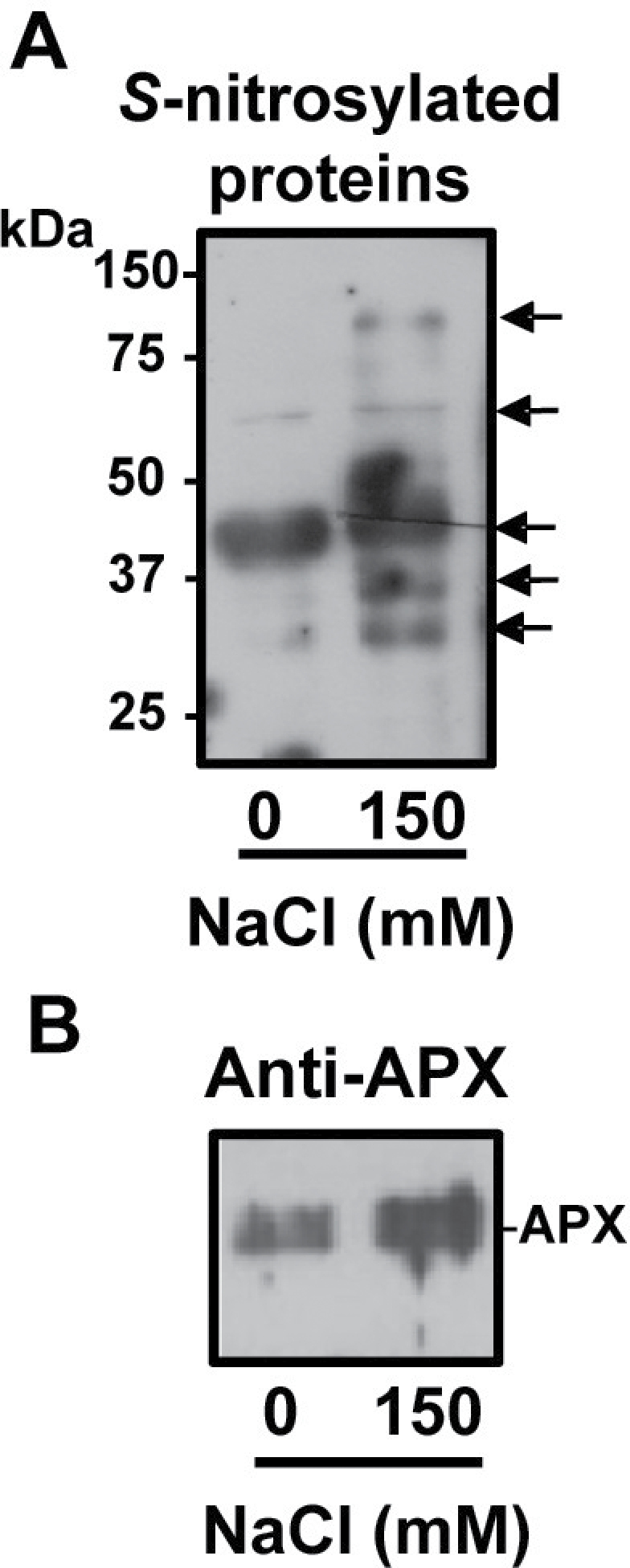
Post-translational modifications (PTMs) mediated by nitric oxide (NO)-derived molecules have become a new area of research, as they can modulate the function of target proteins. Proteomic data have shown that ascorbate peroxidase (APX) is one of the potential targets of PTMs mediated by NO-derived molecules. Using recombinant pea cytosolic APX, the impact of peroxynitrite (ONOO-) and S-nitrosoglutathione (GSNO), which are known to mediate protein nitration and S-nitrosylation processes, respectively, was analysed. While peroxynitrite inhibits APX activity, GSNO enhances its enzymatic activity. Mass spectrometric analysis of the nitrated APX enabled the determination that Tyr5 and Tyr235 were exclusively nitrated to 3-nitrotyrosine by peroxynitrite. Residue Cys32 was identified by the biotin switch method as S-nitrosylated. The location of these residues on the structure of pea APX reveals that Tyr235 is found at the bottom of the pocket where the haem group is enclosed, whereas Cys32 is at the ascorbate binding site. Pea plants grown under saline (150 mM NaCl) stress showed an enhancement of both APX activity and S-nitrosylated APX, as well as an increase of H2O2, NO, and S-nitrosothiol (SNO) content that can justify the induction of the APX activity. The results provide new insight into the molecular mechanism of the regulation of APX which can be both inactivated by irreversible nitration and activated by reversible S-nitrosylation.

Keywords: Ascorbate peroxidase; S-nitrosoglutathione; S-nitrosylation; nitration; nitric oxide; peroxynitrite; reactive nitrogen species; salinity stress..


Figures

Fig. 1.
Fig. 1.
SDS–PAGE analysis of the purification of the recombinant cytosolic ascorbate peroxidase (APX). The gel was stained with Coomasie blue. M, molecular markers; I, total protein in induced culture; SF, soluble fraction; IF, insoluble fraction; FT, flow-through; W, wash; E1–E8, elution fractions.
Fig. 2.
Fig. 2.
Effect of nitration (A and B) and S-nitrosylation on recombinant ascorbate peroxidase (APX) (C–E). (A) Effect of SIN-1 (peroxynitrite donor) on recombinant APX activity. (B) Representative immunoblot showing the grade of tyrosine nitration of APX treated with different concentrations of SIN-1 and detected with an antibody against 3-nitrotyrosine (dilution 1:2500). A 5 μg aliquot of protein was used per line. (C) Effect of S-nitrosoglutathione (GSNO). (D) Effect of glutathione (GSH). (E) S-nitrosylation of recombinant pea APX. A 5 μg aliquot of purified recombinant APX was treated with 2mM GSH and 2mM GSNO and was subjected to the biotin switch method. Control treatments were done with water (lane 1) and 2mM GSH (lane 2). Additionally, APX was S-nitrosylated with 2mM GSNO and reduced again with 50mM DTT (lane 4). Furthermore, GSNO-treated APX underwent the biotin switch method without ascorbate (lane 5). Proteins were separated under non-reducing conditions by SDS–PAGE and blotted onto a PVDF membrane. Biotinylated proteins were detected using anti-biotin antibodies. Ponceau red staining demonstrated equal loading.
Fig. 3.
Fig. 3.
Comparison of the nitrated (top) and unmodified (bottom) MS/MS spectra of the identified peptides from pea APX in the corresponding panels: (A) YAADEDVFFADY*AEAHLK. (B) SY*PTVSPDYQK. Peptide fragment ions are indicated by ‘b’ if the charge is retained on the N-terminus and by ‘y’ if the charge is maintained on the C-terminus. The subscript indicates the number of amino acid residues in the considered fragment from either the N-terminus or the C-terminus. The superscript indicates the charge (1+ or 2+) of the backbone fragmentation.
Fig. 4.
Fig. 4.
(A) Structure of homodimeric pea APX (PDB ID: 1apx). Residues identified as a target of tyrosine nitration and S-nitrosylation are shown as space filling. (B) The haem group is enclosed in a pocket with two channels to the exterior. (C) The view along the upper channel reveals that Y235 is at the bottom of the pocket. (D) C32 is located at the ascorbate binding site in the vicinity of the side channel. (This figure is available in colour at JXB online.)
Fig. 5.
Fig. 5.
Lipid peroxidation, hydrogen peroxide, and ascorbate peroxidase (APX) activity, and representative images illustrating the CLSM detection and visualization of NO and S-nitrosothiols (SNOs) in leaves of pea plants exposed to 150mM NaCl. (A) Malondialdehyde (MDA) content. (B) Hydrogen peroxide content. (C) APX activity and western blotting analysis using an antibody against cucumber APX (dilution 1:3000). Lanes 1 and 2 correspond to leaf extracts of pea control plants and plants exposed to 150mM NaCl, respectively. (D) Detection of NO in a leaf cross-section of pea control plants (0mM NaCl). (E) Detection of NO in a leaf cross-section of pea plants exposed to 150mM NaCl. (F) Detection of SNOs in a leaf cross-section of pea control plants (0mM NaCl). (G) Detection of SNOs in a leaf cross-section of pea plants exposed to 150mM NaCl. NO and SNOs were detected with the fluorescent dyes DAF-FM DA and Alexa Fluor (AL) 488 Hg-link reagents, respectively, as described in the Materials and methods. The chlorophyll autofluorescence is shown. Adaxial epidermis (E1), abaxial epidermis (E2), main vein (V), palisade mesophyll (Pm), and spongy mesophyll (Sm). Bar=300 μm. Data are means ±SEM of at least three replicates. *Differences from control values were significant at P < 0.05. (This figure is available in colour at JXB online.)
Fig. 6.
Fig. 6.
Total S-nitrosylated proteins and S-nitrosylated APX in leaves of pea plants under salinity stress conditions. (A) Detection of total S-nitrosylated proteins from leaves of pea plant controls and those exposed to 150mM NaCl. S-nitrosylated proteins were separated under non-reducing conditions by 12% SDS–PAGE and blotted onto a PVDF membrane. Biotinylated proteins were detected using anti-biotin antibodies as described in the Materials and methods. (B) Immunoblot of total S-nitrosylated proteins of leaves of pea plant controls and those exposed to 150mM NaCl probed with a polyclonal antibody against cucumber APX (dilution 1:3,000). A 5 μg aliquot of protein was used per lane.

References

    1. Álvarez C, Lozano-Juste J, Romero LC, García I, Gotor C, León J. 2011. Inhibition of Arabidopsis O-acetylserine(thiol)lyase A1 by tyrosine nitration. Journal of Biological Chemisty 286, 578–586 - PMC - PubMed
    1. Astier J, Kulik A, Koen E, Besson-Bard A, Bourque S, Jeandroz S, Lamotte O, Wendehenne D. 2012. Protein S-nitrosylation: what’s going on in plants? Free Radical Biology and Medicine 53, 1101–10 - PubMed
    1. Asada K. 1992. Ascorbate peroxidase: a hydrogen peroxide-scavenging enzyme in plants. Physiologia Plantarum 85, 235–241
    1. Bai X, Yang L, Tian M, Chen J, Shi J, Yang Y, Hu X. 2011. Nitric oxide enhances desiccation tolerance of recalcitrant Antiaris toxicaria seeds via protein S-nitrosylation and carbonylation. PLoS One 6, e20714. - PMC - PubMed
    1. Begara-Morales JC, Chaki M, Sánchez-Calvo B, Mata-Pérez C, Leterrier M, Palma JM, Barroso JB, Corpas FJ. 2013a. Protein tyrosine nitration in pea roots during development and senescence. Journal of Experimental Botany 64, 1121–1134 - PMC - PubMed

Publication types

MeSH terms