

doi: 10.1126/science.1241888.
Nonenzymatic template-directed RNA synthesis inside model protocells
Affiliations
- PMID: 24288333
- PMCID: PMC4104020
- DOI: 10.1126/science.1241888
Item in Clipboard
Nonenzymatic template-directed RNA synthesis inside model protocells
Science.
.
Abstract
Efforts to recreate a prebiotically plausible protocell, in which RNA replication occurs within a fatty acid vesicle, have been stalled by the destabilizing effect of Mg(2+) on fatty acid membranes. Here we report that the presence of citrate protects fatty acid membranes from the disruptive effects of high Mg(2+) ion concentrations while allowing RNA copying to proceed, while also protecting single-stranded RNA from Mg(2+)-catalyzed degradation. This combination of properties has allowed us to demonstrate the chemical copying of RNA templates inside fatty acid vesicles, which in turn allows for an increase in copying efficiency by bathing the vesicles in a continuously refreshed solution of activated nucleotides.
Figures
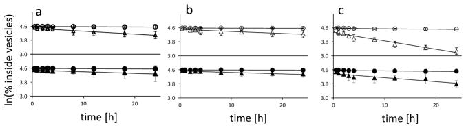
Leakage of a small charged molecule (calcein, open symbols) and a larger 9-mer oligodeoxynucleotide (filled symbols) from fatty acid vesicles. A: oleate vesicles; B: myristoleate: glycerol monomyristoleate 2:1 vesicles; C: decanoate: decanol: glycerol monodecanoate 4:1:1 vesicles. Circles: no MgCl2, no citrate; triangles: 50 mM MgCl2, 200 mM Na+-citrate. The assay used to obtain this data is described in Figure. Lines are linear fits, R2 ≥ 0.97. All experiments were repeated twice, error bars are mean ±2SE.
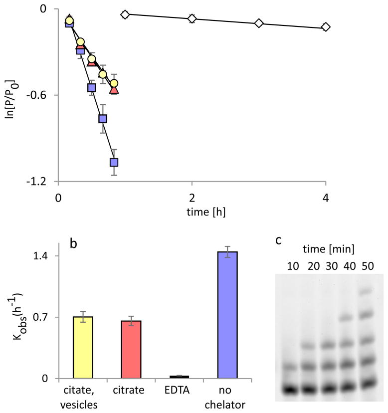
A: Time courses of primer extension on a templating region of four C residues, expressed as fraction of un-extended primer remaining vs. time. Squares: no chelators; triangles: 200 mM Na+-citrate; circles: 200 mM Na+-citrate and 100 mM oleate vesicles; diamonds: 200 mM EDTA. Lines are linear fits, R2 ≥ 0.97; the slope is kobs (h−1). B: Rates of primer extension under the indicated conditions; each kinetic experiment was performed in duplicate and rates are determined as average of the three separate runs. Error bars indicate SEM, n=3. C: Typical PAGE analysis of a template-directed primer extension experiment. Primer extension was carried out in the presence of 200 mM Na+-citrate. For the gel analysis of the reactions used to obtain this data, see Supporting Information Figure S15.
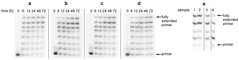
A to D: Primer extension on a templating region of seven C residues. A: control reaction in solution; B: inside oleate vesicles, C: inside myristoleate: glycerol monomyristoleate 2:1 vesicles; D: inside decanoate: decanol: glycerol monodecanoate 4:1:1 vesicles. E: Extension of labeled RNA primer annealed to a mixed base template, templating region sequence GCCG. Sample 1: reaction inside myristoleate: glycerol monomyristoleate 2:1 vesicles, sample 2: reaction inside decanoate: decanol: glycerol monodecanoate 4:1:1 vesicles, both sample 1 and 2 reactions preformed inside liposome dialyzer (see Supplementary Materials for the description of the liposome dialyzer) with a total of 13 buffer exchanges. Sample 3: control reaction in solution with daily addition of fresh portion of activated monomers, without removing the hydrolyzed monomers. Sample 4: control reaction in solution, without addition of fresh monomer.
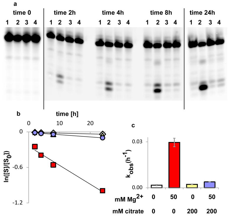
A: PAGE analysis of cleavage of a DNA oligonucleotide at the site of a single internal ribonucleotide, at indicated time points; lanes 1: no Mg2+, no citrate; lanes 2: 50 mM Mg2+, no citrate; lanes 3: no Mg2+, 200 mM citrate; lanes 4: 50 mM Mg2+, 200 mM citrate. B: Quantitation of strand cleavage, expressed as fraction of intact substrate over total substrate vs. time: diamonds: no Mg2+, no citrate; triangles: no Mg2+, 200 mM citrate; circles: 50 mM Mg2+, 200 mM citrate; squares 50 mM Mg2+, no citrate. Lines are linear fits, R2 ≥ 0.97. C: Rates of strand cleavage at the ribo linkage with and without Mg2+ and citrate. Error bars indicate SEM, n=3.
References
-
- Gilbert W. Origin of life: The RNA world. Nature. 1986;319:618.
-
- Joyce GF. RNA evolution and the origins of life. Nature. 1989;338:217. - PubMed
-
- Gebicki JM, Hicks M. Ufasomes are stable particles surrounded by unsaturated fatty acid membranes. Nature. 1973;243:232. - PubMed
-
- Deamer DW, Dworkin JP. Chemistry and physics of primitive membranes. Top Curr Chem. 2005;1
-
- Szostak JW. The eightfold path to non-enzymatic RNA replication. J Sys Chem. 2012;3
Publication types
MeSH terms
Substances
Grants and funding
LinkOut - more resources
Full Text Sources
Other Literature Sources
