. 2013:2013:817674.
doi: 10.1155/2013/817674. Epub 2013 Oct 31.

Subtoxic Levels of Apigenin Inhibit Expression and Secretion of VEGF by Uveal Melanoma Cells via Suppression of ERK1/2 and PI3K/Akt Pathways

Affiliations

Subtoxic Levels of Apigenin Inhibit Expression and Secretion of VEGF by Uveal Melanoma Cells via Suppression of ERK1/2 and PI3K/Akt Pathways

Shih-Chun Chao et al. Evid Based Complement Alternat Med. 2013.

Abstract

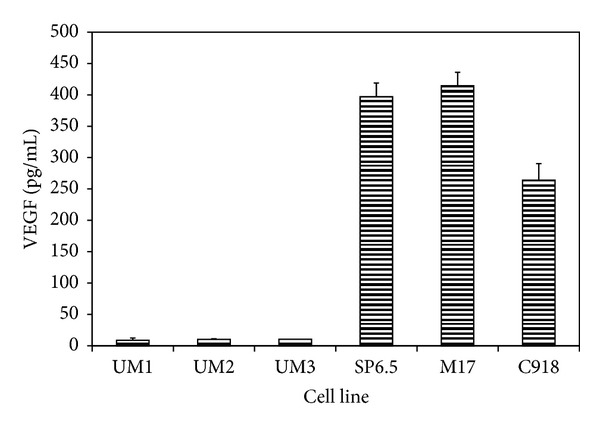
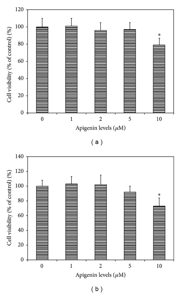
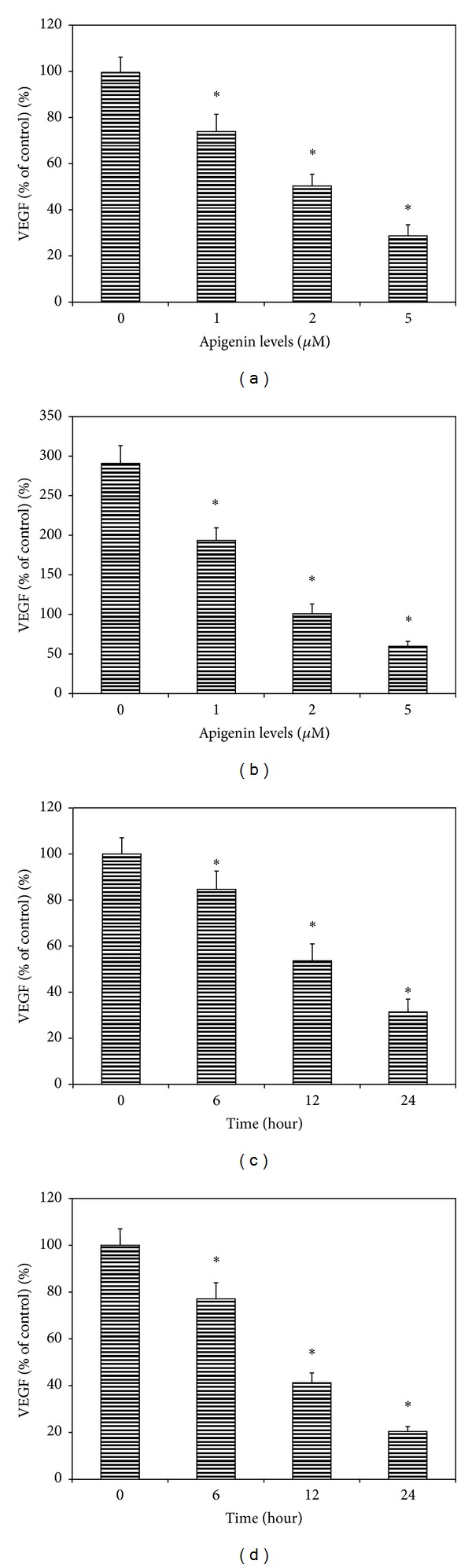
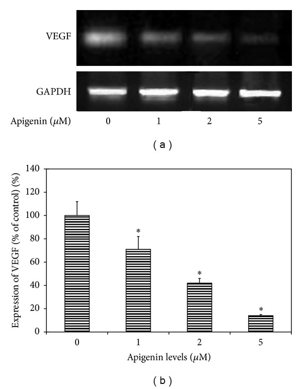
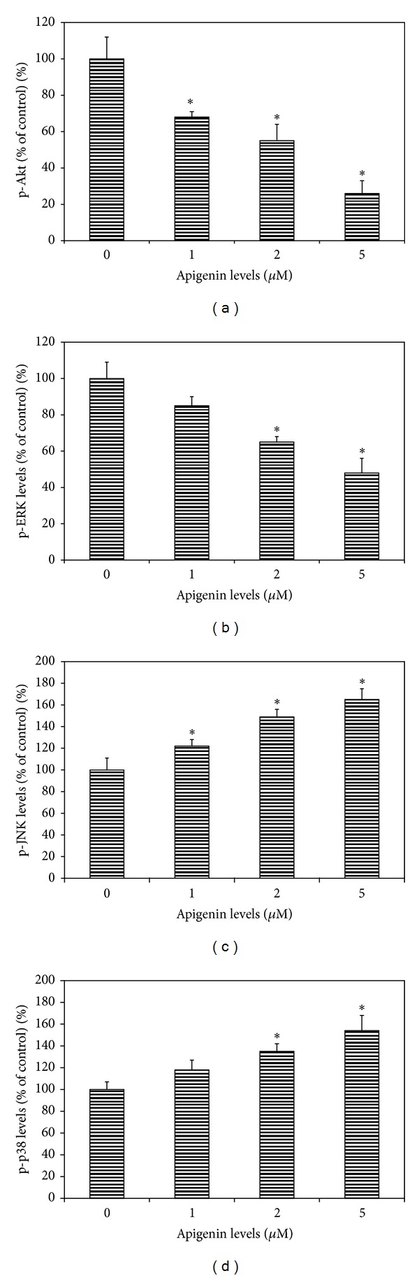
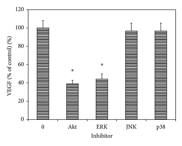
The effects of apigenin on the expression of VEGF in uveal melanoma cells have not been reported. We studied this effect and relevant signaling pathways in two human uveal melanoma cell lines (SP6.5 and C918). ELISA assay revealed that the constitutive secretion of VEGF by uveal melanoma cells was 21-fold higher than that in normal uveal melanocytes. Apigenin at subtoxic levels (1-5 μ M) significantly suppressed the secretion of VEGF in a dose- and time-dependent manner in melanoma cells. VEGF levels in the conditioned culture media from SP6.5 and C918 cell lines treated with 5 μ M apigenin for 24 h reduced to 29% and 21% of those in cells not treated with apigenin, respectively. RT-PCR analysis found that apigenin also decreased the expression of VEGF mRNA in melanoma cells. ELISA study of various signal pathways showed that apigenin significantly decreased phosphorylated Akt and ERK1/2 but increased phosphorylated JNK1/2 and p38 MAPK levels in melanoma cells. PI3K/Akt or ERK1/2 inhibitors significantly decreased, but JNK1/2 and p38 MAPK inhibitors did not influence the secretion of VEGF by melanoma cells, suggesting that apigenin suppresses the secretion of VEGF mainly through the inhibition of PI3K/Akt and ERK1/2 pathways.


Figures

Figure 1
Figure 1
Constitutive secretion of VEGF by human uveal melanoma cells and normal uveal melanocytes. Three different primary cultures of uveal melanocytes (UM1-3) and uveal melanoma cells from 3 different cell lines (SP6.5, M17, and C918) were plated into 24-well plates. After 24 h, culture medium was removed and cultures were washed by PBS. Serum-free culture medium was added and cultured for 24 h. Conditioned media were collected, and the amount of VEGF protein was determined by using the human VEGF Quantikine ELISA kit. VEGF levels in conditioned culture medium were expressed as pg/mL. VEGF levels in culture medium from melanoma cells were significantly higher than that from normal melanocytes (P < 0.05). Data are mean ± SD (n = 3).
Figure 2
Figure 2
Dose effect of apigenin on cell viability of uveal melanoma cells. Human uveal melanoma cells SP6.5 (a) and C918 (b) were seeded into 96-well plates and treated with apigenin at various doses for 48 h, and cell viability was determined by MTT assay (see Methods). Apigenin at 1–5 μM did not affect, whereas 10 μM apigenin significantly decreased the cell viability of uveal melanoma cells. Data are mean ± SD (n = 3). *P < 0.05, versus control (cells cultured without apigenin).
Figure 3
Figure 3
Dose- and time-effects of apigenin on the secretion of VEGF by human uveal melanoma cells. Cells were plated into 24-well plates. In the dose-effect study, apigenin at 0, 1, 2 and 5 μM was added to the culture and incubated for 24 h, (a) SP6.5 cell line and (b) C918 cell line. In the time-effect study, apigenin at 5.0 μM was added and conditioned medium was collected 6, 12 and 24 h later, (c) SP6.5 and (d) C918. The amount of VEGF protein in the conditioned medium was determined by using the human VEGF Quantikine ELISA kit. VEGF levels in the conditioned culture medium were expressed as the percentages of the controls. Apigenin significantly reduced the secretion of VEGF by uveal melanoma cells in a dose- and time-dependent manner. Data are mean ± SD (n = 3). *P < 0.05, versus control (cells cultured without apigenin).
Figure 4
Figure 4
RT-PCR analysis for the effects of apigenin on the expression of VEGF mRNA in uveal melanoma cells. Representative RT-PCR profiles from three experiments showed the mRNA expressions of VEGF by uveal melanoma cells (SP6.5 cell line) exposed to apigenin at 0, 1, 2, and 5 μM (a). After 6 h of culture with or without apigenin, cells were collected, mRNA was extracted, and RT-PCR analysis was performed as described in the text. GAPDH was used as an internal loading control. Apigenin inhibited the expression of VEGF mRNA in uveal melanoma cells in a dose-dependent manner (b).
Figure 5
Figure 5
Dose effects of apigenin on phosphorylated Akt, ERK, JNK, and p38 MAPK levels in cultured uveal melanoma cells. Cells (SP6.5) were plated into 24-well plates. After 24 h incubation, apigenin at 0, 1, 2, and 5 μM was added to the medium. Cells were collected 60 min later. The amount of phosphorylated Akt (p-Akt), ERK1/2 (p-ERK), JNK1/2 (p-JNK), and p38 MAPK (p-p38) in cell lysates was measured using the relevant ELISA kits and was expressed as the percentages of the controls (cells cultured without apigenin). Apigenin significantly decreased p-Akt (a) and p-ERK levels (b) and increased p-JNK (c) and p-p38 (d) levels in cell lysates in a dose-dependent manner. Data are mean ± SD (n = 3). *P < 0.05, versus control.
Figure 6
Figure 6
Effects of Akt and MAPK inhibitors on the constitutive secretion of VEGF by uveal melanoma cells. Cells were plated into 24-well plates. Various MAPK and Akt inhibitors were added to the medium separately, including LY294002 (PI3K/Akt inhibitor), UO1026 (ERK inhibitor), SP600125 (JNK inhibitor), and SB203580 (p38 MAPK inhibitor) at a final concentration of 10 μM. After 24 h incubation, culture medium was collected, the VEGF levels were measured with human VEGF Quantikine ELISA kit and expressed as the percentages of the controls (cells cultured without any signal inhibitors). Akt and ERK inhibitors significantly decreased, whereas JNK and p38 inhibitors did not affect the VEGF levels in the conditioned culture medium from uveal melanoma cells. Data are mean ± SD (n = 3). *P < 0.05, versus control (cells cultured without inhibitor).

Similar articles

Cited by

References

    1. Hu DN, Yu GP, McCormick SA, Schneider S, Finger PT. Population-based incidence of uveal melanoma in various races and ethnic groups. American Journal of Ophthalmology. 2005;140(4):612–617. - PubMed
    1. Kujala E, Mäkitie T, Kivelä T. Very long-term prognosis of patients with malignant uveal melanoma. Investigative Ophthalmology and Visual Science. 2003;44(11):4651–4659. - PubMed
    1. Augsburger JJ, Corrêa ZM, Shaikh AH. Effectiveness of treatments for metastatic uveal melanoma. American Journal of Ophthalmology. 2009;148(1):119–127. - PubMed
    1. Xie K, Wei D, Shi Q, Huang S. Constitutive and inducible expression and regulation of vascular endothelial growth factor. Cytokine and Growth Factor Reviews. 2004;15(5):297–324. - PubMed
    1. Loureiro RMB, D’Amore PA. Transcriptional regulation of vascular endothelial growth factor in cancer. Cytokine and Growth Factor Reviews. 2005;16(1):77–89. - PubMed

LinkOut - more resources