Review
. 2013:2013:734509.
doi: 10.1155/2013/734509. Epub 2013 Oct 28.

Impact of endothelial microparticles on coagulation, inflammation, and angiogenesis in age-related vascular diseases

Affiliations
Review

Impact of endothelial microparticles on coagulation, inflammation, and angiogenesis in age-related vascular diseases

Margaret Markiewicz et al. J Aging Res. 2013.

Abstract

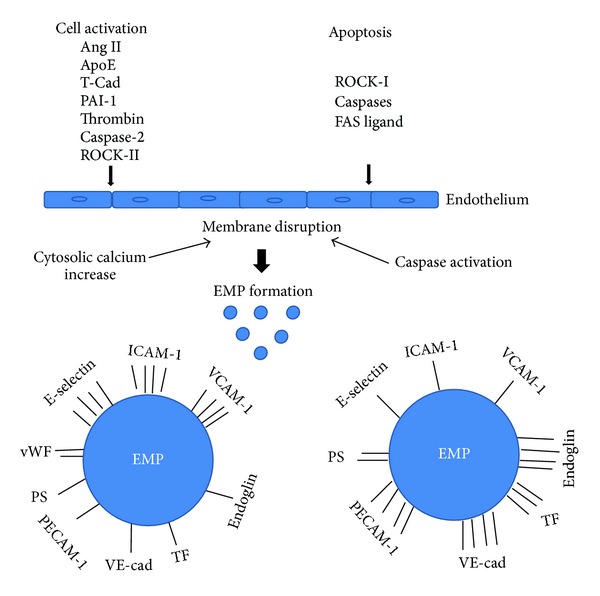
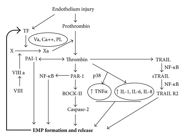
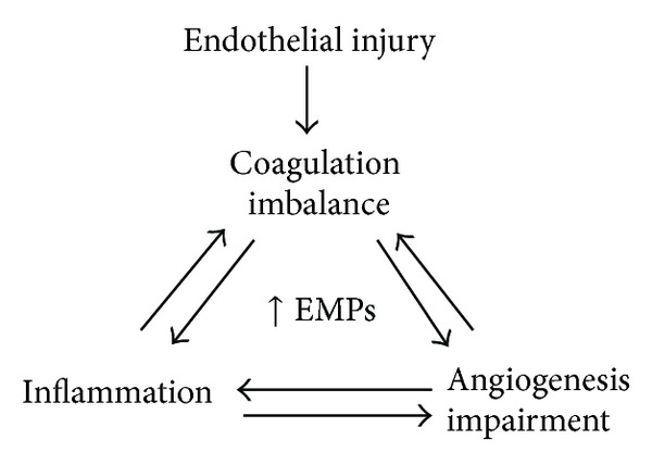
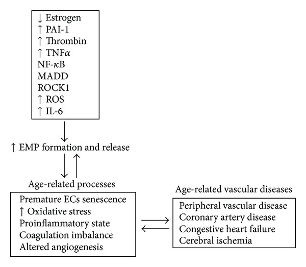
Endothelial microparticles (EMPs) are complex vesicular structures that originate from plasma membranes of activated or apoptotic endothelial cells. EMPs play a significant role in vascular function by altering the processes of inflammation, coagulation, and angiogenesis, and they are key players in the pathogenesis of several vascular diseases. Circulating EMPs are increased in many age-related vascular diseases such as coronary artery disease, peripheral vascular disease, cerebral ischemia, and congestive heart failure. Their elevation in plasma has been considered as both a biomarker and bioactive effector of vascular damage and a target for vascular diseases. This review focuses on the pleiotropic roles of EMPs and the mechanisms that trigger their formation, particularly the involvement of decreased estrogen levels, thrombin, and PAI-1 as major factors that induce EMPs in age-related vascular diseases.


Figures

Figure 1
Figure 1
Differences in antigen expression of EMPs derived from activation versus apoptosis of endothelial cells. Endothelial cell activators, angiotensin II (Ang II), apoliprotein E (ApoE), T-cadherin (T-Cad), plasminogen activator inhibitor-1 (PAI-1), and thrombin, cause cytosolic calcium increase which leads to endothelial cell membrane disruption. Caspase-2 activates ROCK-II independently of cell death [45]. Apoptosis inducers including Rho activated kinase (ROCK-I), caspases, and FAS ligand activate caspases and cause membrane disruption in endothelial cells. Both activators of cell activation and apoptosis lead to vesiculation and EMP generation. Bars represent level of antigen on the surface of the EMP. 4 bars: high level of antigen; 1 bar: low level of antigen. EMP antigens: E-selectin, intracellular adhesion molecule-1 (ICAM-1), vascular cell adhesion molecule-1 (VCAM-1), platelet cell adhesion molecule-1 (PECAM-1), endoglin, vascular endothelial-cadherin (VE-cadherin), tissue factor (TF), phospholipid (PS), and von Willebrand factor (vWf). This figure was prepared based on [, –40, 42, 45].
Figure 2
Figure 2
Signaling pathways involved in thrombin induced EMP formation. EMPs carry TF, the main initiator of the extrinsic pathway of the coagulation cascade. Cofactor VIIIa known as a von Willebrand factor activates factor X to Xa in the presence of factor Va, calcium, and phospholipids (PL), which results in the generation of thrombin. Thrombin induces tumor necrosis factor alpha (TNFα) via p38 (mitogen activated protein kinase) and interleukins 1, 6, and 8 (Il-1, IL-6, and IL-8), which both lead to formation of EMPs. Thrombin, via proteolytically activated receptor-1 (PAR-1), induces nuclear factor kappa B (NF-κB), which directly induces EMP formation. PAR-1 also induces Rho kinase (ROCK- II) which activates caspase-2 leading to EMPs formation. Thrombin via (tumor necrosis factor related apoptosis inducing ligand) TRAIL activates sTRAIL, which is synthesized to TRAIL R2. This process requires the activation of NF-κB.
Figure 3
Figure 3
EMPs link inflammation, coagulation, and angiogenesis.
Figure 4
Figure 4
Factors contributing to EMP formation in age-related vascular diseases.

References

    1. Liu W, Reinmuth N, Stoeltzing O, et al. Antiangiogenic therapy targeting factors that enhance endothelial cell survival. Seminars in Oncology. 2002;29(3):96–103. - PubMed
    1. Endemann DH, Schiffrin EL. Endothelial dysfunction. Journal of the American Society of Nephrology. 2004;15(8):1983–1992. - PubMed
    1. Burger D, Montezano AC, Nishigaki N, He Y, Carter A, Touyz RM. Endothelial microparticle formation by angiotensin II is mediated via ang II receptor type I/NADPH Oxidase/rho kinase pathways targeted to lipid rafts. Arteriosclerosis, Thrombosis, and Vascular Biology. 2011;31(8):1898–1907. - PubMed
    1. Leroyer AS, Anfosso F, Lacroix R, et al. Endothelial-derived microparticles: biological conveyors at the crossroad of inflammation, thrombosis and angiogenesis. Thrombosis and Haemostasis. 2010;104(3):456–463. - PubMed
    1. Owens AP, MacKman N. Microparticles in hemostasis and thrombosis. Circulation Research. 2011;108(10):1284–1297. - PMC - PubMed