. 2013:2013:843897.
doi: 10.1155/2013/843897. Epub 2013 Oct 31.

Inhibition of Angiogenesis In Vitro by Chebulagic Acid: A COX-LOX Dual Inhibitor

Affiliations

Inhibition of Angiogenesis In Vitro by Chebulagic Acid: A COX-LOX Dual Inhibitor

A P Athira et al. Int J Vasc Med. 2013.

Abstract

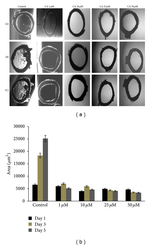
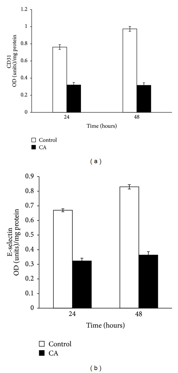
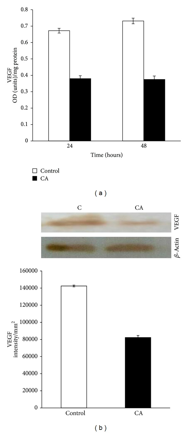
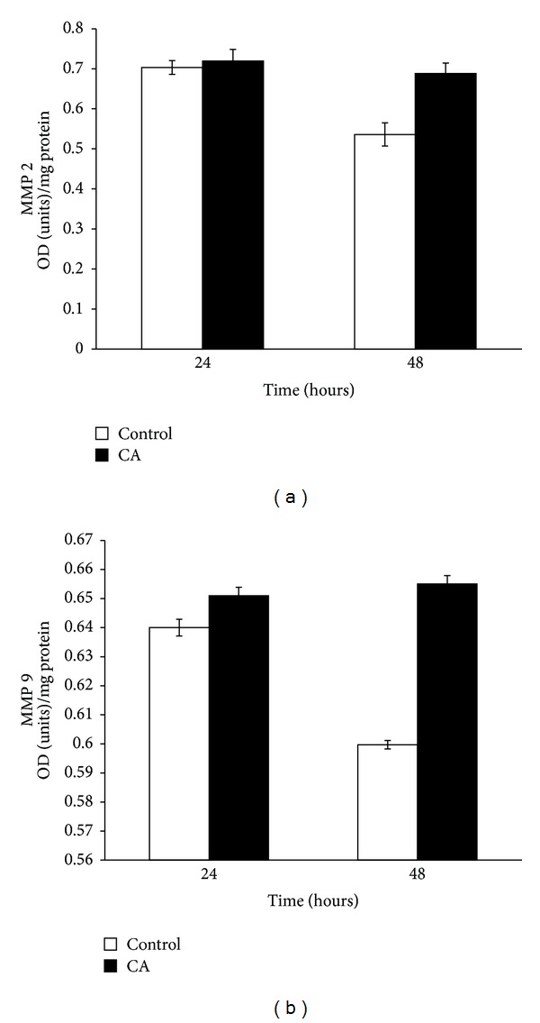
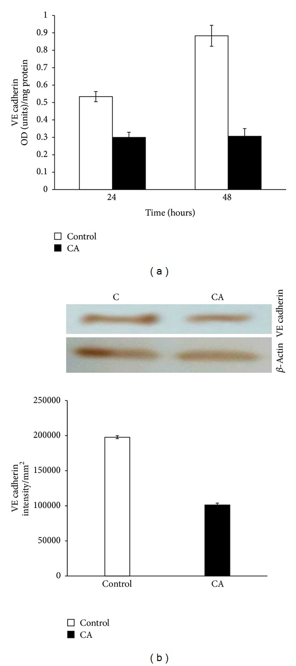
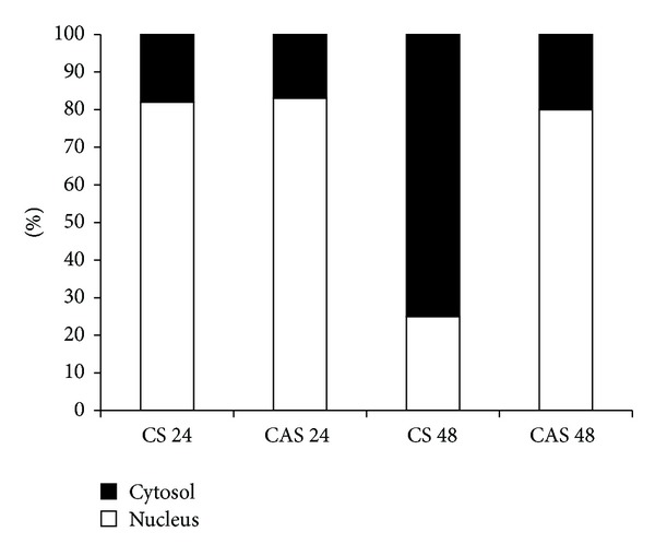
Angiogenesis is a crucial step in the growth of cancer and its metastasis. It is regulated by several endogenous factors which may stimulate or inhibit the new blood vessel growth. Besides these endogenous factors, several exogenous factors including some natural compounds are known to modulate angiogenesis. Angiogenesis being a potential target for drugs against a number of pathological conditions, search for compounds from natural sources that can affect angiogenesis is of great interest. The objective of our present study was to understand the effect of chebulagic acid, a COX-LOX dual inhibitor isolated from the fruits of Terminalia chebula Retz., on angiogenesis. The model systems used were rat aortic rings and human umbilical vein endothelial cells. The results showed that chebulagic acid exerts an antiangiogenic effect. This was evidenced from decreased sprouting in rat aortic rings and decrease in biochemical markers in endothelial cells treated with chebulagic acid. It downregulated the production of CD31, E-selectin, and vascular endothelial growth factor in human umbilical vein endothelial cells in culture (HUVEC). Further studies to understand the molecular mechanism of action of chebulagic acid revealed that CA exerts its anti angiogenic effect by modulating VE cadherin-β catenin signalling in human umbilical vein endothelial cells.


Figures

Figure 1
Figure 1
(a) Effect of chebulagic acid on sprouting of rat aortic rings. Rat aortic ring explants were maintained in culture in the presence of 10% fetal bovine serum (FBS). Different concentrations of chebulagic acid (1 μM, 10 μM, 25 μM, and 50 μM) were supplemented to the medium and was monitored sprouting, under a microscope at different time intervals. The morphological changes were visualized and photographed under a microscope (×4) on the 1st (A), 3rd (B), and 5th (C) days. Each set was done in replicates, and the microphotographs from a representative experiment are given. (b) The sprouts were quantitated using QWIN software (Leica) and expressed in area in µm2. Values given are the average of five experiments ± SEM, statistically significant compared to control (P < 0.05).
Figure 2
Figure 2
Effect of CA on the expression of CD31 and E-selectin. HUVECs were maintained in culture in MCDB 131 medium containing 10% FBS supplemented with 25 μM CA for 48 hours. The levels of cell-associated CD31 (a) and E-selectin secreted into the medium (b) at 24 and 48 hours were estimated by ELISA using anti-CD31 (1 : 1000) and anti-E-selectin (1 : 1000) antibody, respectively. Values given are the average of duplicate analysis of 5 experiments ± SEM (P < 0.05).
Figure 3
Figure 3
Effect of CA on the expression of VEGF. HUVECs were maintained in culture in MCDB 131 medium containing 10% FBS supplemented with 25 μM CA for 48 hours. (a) The level of VEGF secreted into the medium was estimated using anti-VEGF antibody. Values given are the average of duplicate analysis of 5 experiments ± SEM (P < 0.05). (b) Western blot to analyze the production of VEGF by control cells (C) and chebulagic acid treated cells (CA) at 48 hours. The intensity of the immunoblotted bands was measured, normalised to intensity of internal control actin, and expressed in intensity units/mm2.
Figure 4
Figure 4
HUVECs maintained in culture in MCDB 131 containing 10% FBS for 48 hours were treated with 25 μM CA. The level of MMP 2 and MMP 9 in the medium was estimated by ELISA using anti-MMP 2 (1 : 1000) (a) and MMP 9 (1 : 1000) (b) antibody, respectively. Values given are the average of duplicate analysis of 5 experiments ± SEM (P < 0.05).
Figure 5
Figure 5
Effect of CA on the expression of VE cadherin. HUVECs were maintained in culture in MCDB 131 medium containing 10% FBS supplemented with 25 μM CA for 48 hours. (a) The level of VE cadherin in the cell layer was estimated using anti-VE cadherin antibody. Values given are the average of duplicate analysis of 5 experiments ± SEM (P < 0.05). (b) Western blot to analyze the production of VE cadherin by control cells (C) and chebulagic acid treated cells (CA) at 48 hours. The intensity of the immunoblotted bands was measured, normalised to that of internal control, and expressed in intensity units/mm2. Values given are mean of 5 experiments ± SEM.
Figure 6
Figure 6
Effect of CA on the distribution of β catenin. HUVECs maintained in culture in MCDB 131 medium containing 10% FBS were supplemented with 25 μM CA. The level of β catenin in the nucleus and cytosol at 24 and 48 hours was estimated by ELISA using anti-β catenin antibody (1 : 1000) and expressed as percent of total. CS 24 and CS 48: control at 24 and 48 hours. CAS 24 and CAS 48: chebulagic acid at 24 and 48 hours.

References

    1. Hanahan D, Weinberg RA. Hallmarks of cancer: the next generation. Cell. 2011;144(5):646–674. - PubMed
    1. Carmeliet P. Blood vessels and nerves: common signals, pathways and diseases. Nature Reviews Genetics. 2003;4(9):710–720. - PubMed
    1. Hanahan D, Folkman J. Patterns and emerging mechanisms of the angiogenic switch during tumorigenesis. Cell. 1996;86(3):353–364. - PubMed
    1. Bouck N. Tumor angiogenesis: the role of oncogenes and tumor suppressor genes. Cancer Cells. 1990;2(6):179–185. - PubMed
    1. Reddy DB, Reddanna P. Chebulagic acid (CA) attenuates LPS-induced inflammation by suppressing NF-κB and MAPK activation in RAW 264.7 macrophages. Biochemical and Biophysical Research Communications. 2009;381(1):112–117. - PubMed

LinkOut - more resources