Review
. 2013 Oct 31:2013:134858.
doi: 10.1155/2013/134858. eCollection 2013.

The Afferent Visual Pathway: Designing a Structural-Functional Paradigm of Multiple Sclerosis

Affiliations
Review

The Afferent Visual Pathway: Designing a Structural-Functional Paradigm of Multiple Sclerosis

Fiona Costello. ISRN Neurol. .

Abstract

Multiple sclerosis (MS) is a disease of the central nervous system (CNS) believed to arise from a dysfunctional immune-mediated response in a genetically susceptible host. The actual cause of MS is not known, and there is ongoing debate about whether this CNS disorder is predominantly an inflammatory versus a degenerative condition. The afferent visual pathway (AVP) is frequently involved in MS, such that one in every five individuals affected presents with acute optic neuritis (ON). As a functionally eloquent system, the AVP is amenable to interrogation with highly reliable and reproducible tests that can be used to define a structural-functional paradigm of CNS injury. The AVP has numerous unique advantages as a clinical model of MS. In this review, the parameters and merits of the AVP model are highlighted. Moreover, the roles the AVP model may play in elucidating mechanisms of brain injury and repair in MS are described.


Figures

Figure 1
Figure 1
A schematic diagram of the afferent visual pathway.
Figure 2
Figure 2
(a) A fundus view of the optic nerve and macula. (b) A fundus view of a swollen optic nerve with associated macular edema in a patient with neuroretinitis.
Figure 3
Figure 3
The topographic arrangement of the retinal nerve fiber layer indicating the optic nerve and macula. Nasal RNFL fibers radiate into the optic disc like the spokes of a wheel, whereas the large temporal fibers sweep superiorly and inferiorly around the central area of the retina from the periphery. Small densely packed fibers radiate directly to the mid-temporal optic disc edge from the fovea, making up the papillomacular bundle (image provided by Dr. Kathleen Digre).
Figure 4
Figure 4
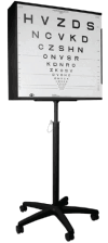
Early Treatment Diabetic Retinopathy Study (ETDRS) chart (image provided by Dr. Laura Balcer).
Figure 5
Figure 5
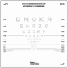
Low-contrast letter acuity chart (image provided by Dr. Laura Balcer).
Figure 6
Figure 6
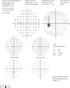
Humphrey perimetry 30-2 threshold testing of the left eye showing the normal physiological blind spot (arrow).
Figure 7
Figure 7
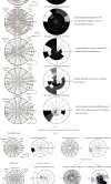
(a) Multifocal VEP amplitude chages after acute optic neuritis. (b) Multifocal VEP showing an example of complete amplitude recovery but significant latency delay (Image provided by Dr. Alexandr Klistorner).
Figure 8
Figure 8
OCT line scan of the macula with consensus layer nomenclature labeling of layers and zones (SPECTRALIS® image courtesy of Heidelberg Engineering).

References

    1. Compston A., Coles A. Multiple sclerosis. The Lancet . 2008;372(9648):1502–1517. doi: 10.1016/S0140-6736(08)61620-7. - DOI - PubMed
    1. Racke M. K. The potassium channel KIR4.1 a potential autoantigen in MS. Nature Reviews Neurology . 2012 doi: 10.1038/nneurol.2012.193. - DOI - PubMed
    1. Poser C. M., Paty D. W., Scheinberg L. New diagnostic criteria for multiple sclerosis: guidelines for research protocols. Annals of Neurology . 1983;13(3):227–231. - PubMed
    1. Lublin F. D., Reingold S. C. Defining the clinical course of multiple sclerosis: results of an international survey. Neurology . 1996;46(4):907–911. - PubMed
    1. McDonald W. I., Compston A., Edan G., et al. Recommended diagnostic criteria for multiple sclerosis: guidelines from the International Panel on the Diagnosis of Multiple Sclerosis. Annals of Neurology . 2001;50(1):121–127. doi: 10.1002/ana.1032. - DOI - PubMed

LinkOut - more resources