

Interstitial lung disease associated with Equine Infectious Anemia Virus infection in horses
- PMID: 24289102
- PMCID: PMC3879148
- DOI: 10.1186/1297-9716-44-113
Interstitial lung disease associated with Equine Infectious Anemia Virus infection in horses
Abstract
EIA (Equine Infectious Anemia) is a blood-borne disease primarily transmitted by haematophagous insects or needle punctures. Other routes of transmission have been poorly explored. We evaluated the potential of EIAV (Equine Infectious Anemia Virus) to induce pulmonary lesions in naturally infected equids. Lungs from 77 EIAV seropositive horses have been collected in Romania and France. Three types of lesions have been scored on paraffin-embedded lungs: lymphocyte infiltration, bronchiolar inflammation, and thickness of the alveolar septa. Expression of the p26 EIAV capsid (CA) protein has been evaluated by immunostaining. Compared to EIAV-negative horses, 52% of the EIAV-positive horses displayed a mild inflammation around the bronchioles, 22% had a moderate inflammation with inflammatory cells inside the wall and epithelial bronchiolar hyperplasia and 6.5% had a moderate to severe inflammation, with destruction of the bronchiolar epithelium and accumulation of smooth muscle cells within the pulmonary parenchyma. Changes in the thickness of the alveolar septa were also present. Expression of EIAV capsid has been evidenced in macrophages, endothelial as well as in alveolar and bronchiolar epithelial cells, as determined by their morphology and localization. To summarize, we found lesions of interstitial lung disease similar to that observed during other lentiviral infections such as FIV in cats, SRLV in sheep and goats or HIV in children. The presence of EIAV capsid in lung epithelial cells suggests that EIAV might be responsible for the broncho-interstitial damages observed.
Figures
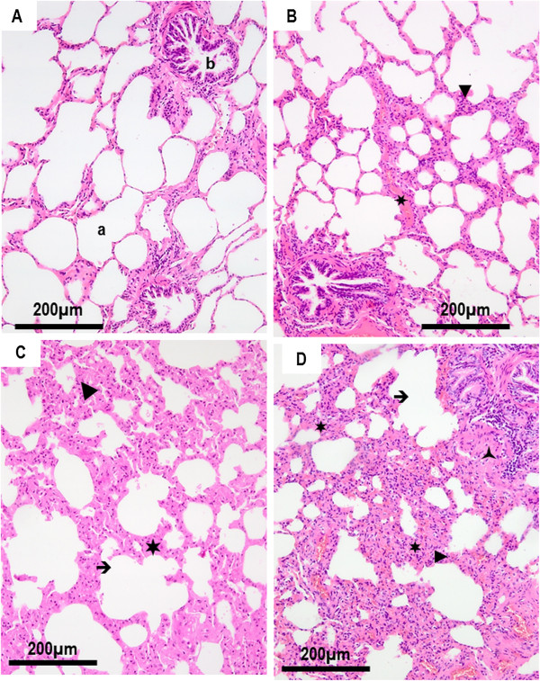
 ) peribronchiolar accumulation of smooth muscle cells; a: alveoli; b: bronchiole.
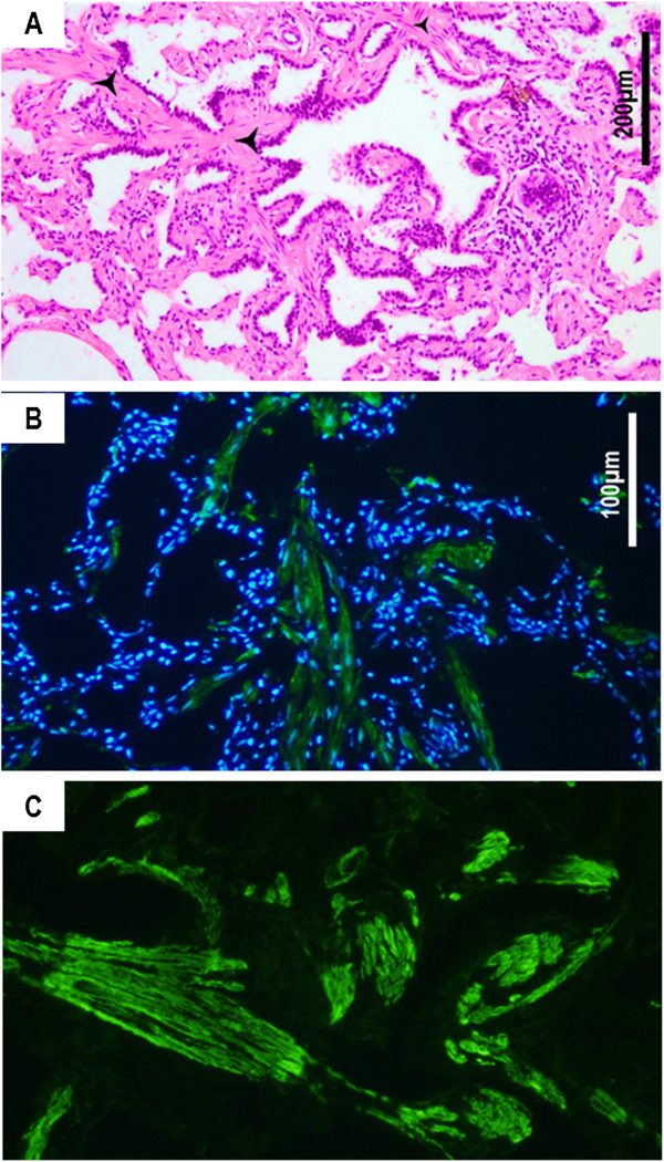
 ) and disrupt the bronchiolar structure. Hematoxylin-eosin staining on paraffin-embedded sections. B. Immunostaining of smooth muscle cells using an anti-αSMA antibody shows the accumulation of fusiform cells (in green) within the lung parenchyma. C. Higher magnification shows organized actin filaments within the infiltrating cells. Nucleus are stained in blue with DAPI.
References
-
- Cappelli K, Capomaccio S, Cook FR, Felicetti M, Marenzoni ML, Coppola G, Verini-Supplizi A, Coletti M, Passamonti F. Molecular detection, epidemiology, and genetic characterization of novel European field isolates of equine infectious anemia virus. J Clin Microbiol. 2011;44:27–33. doi: 10.1128/JCM.01311-10. - DOI - PMC - PubMed
-
- Capomaccio S, Willand ZA, Cook SJ, Issel CJ, Santos EM, Reis JK, Cook RF. Detection, molecular characterization and phylogenetic analysis of full-length equine infectious anemia (EIAV) gag genes isolated from Shackleford Banks wild horses. Vet Microbiol. 2012;44:320–332. doi: 10.1016/j.vetmic.2012.01.015. - DOI - PubMed
Publication types
MeSH terms
Substances
LinkOut - more resources
Full Text Sources
Other Literature Sources
Medical
Miscellaneous
