

En face enhanced-depth swept-source optical coherence tomography features of chronic central serous chorioretinopathy
- PMID: 24289918
- PMCID: PMC3943670
- DOI: 10.1016/j.ophtha.2013.10.014
En face enhanced-depth swept-source optical coherence tomography features of chronic central serous chorioretinopathy
Abstract
Objective: To characterize en face features of the retinal pigment epithelium (RPE) and choroid in eyes with chronic central serous chorioretinopathy (CSCR) using a high-speed, enhanced-depth swept-source optical coherence tomography (SS-OCT) prototype.
Design: Consecutive patients with chronic CSCR were prospectively examined with SS-OCT.
Participants: Fifteen eyes of 13 patients.
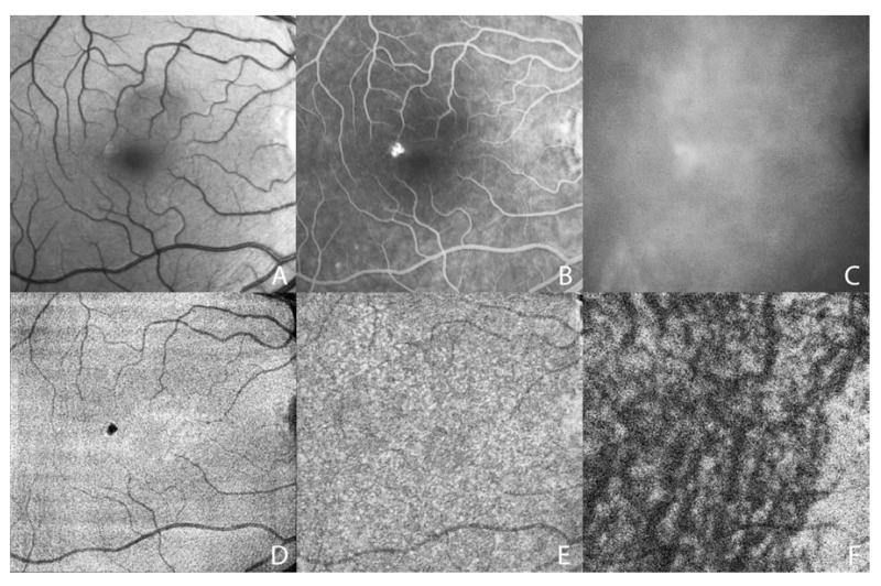
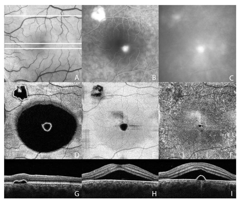
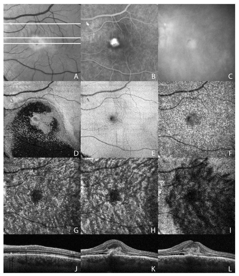
Methods: Three-dimensional 6×6 mm macular cube raster scans were obtained with SS-OCT operating at 1050 nm wavelength and 100000 A-lines/sec with 6 μm axial resolution. Segmentation of the RPE generated a reference surface; en face SS-OCT images of the RPE and choroid were extracted at varying depths every 3.5 μm (1 pixel). Abnormal features were characterized by systematic analysis of multimodal fundus imaging, including color photographs, fundus autofluorescence, fluorescein angiography, and indocyanine-green angiography (ICGA).
Main outcome measures: En face SS-OCT morphology of the RPE and individual choroidal layers.
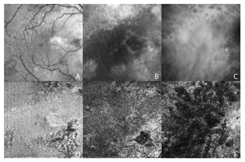
Results: En face SS-OCT imaging at the RPE level revealed absence of signal corresponding to RPE detachment or RPE loss in 15 of 15 (100%) eyes. En face SS-OCT imaging at the choriocapillaris level showed focally enlarged vessels in 8 of 15 eyes (53%). At the level of Sattler's layer, en face SS-OCT documented focal choroidal dilation in 8 of 15 eyes (53%) and diffuse choroidal dilation in 7 of 15 eyes (47%). At the level of Haller's layer, these same features were observed in 3 of 15 eyes (20%) and 12 of 15 eyes (80%), respectively. In all affected eyes, these choroidal vascular abnormalities were seen just below areas of RPE abnormalities. In 2 eyes with secondary choroidal neovascularization (CNV), distinct en face SS-OCT features corresponded to the neovascular lesions.
Conclusions: High-speed, enhanced-depth SS-OCT at 1050 nm wavelength enables the visualization of pathologic features of the RPE and choroid in eyes with chronic CSCR not usually appreciated with standard spectral domain (SD) OCT. En face SS-OCT imaging seems to be a useful tool in the identification of CNV without the use of angiography. This in vivo documentation of the RPE and choroidal vasculature at variable depths may help elucidate the pathophysiology of disease and can contribute to the diagnosis and management of chronic CSCR.
Copyright © 2014 American Academy of Ophthalmology. Published by Elsevier Inc. All rights reserved.
Figures
References
-
- Gass JD. Pathogenesis of disciform detachment of the neuroepithelium. Am J Ophthalmol. 1967;63(suppl):1–139. - PubMed
-
- Yannuzzi LA, Shakin JL, Fisher YL, Altomonte MA. Peripheral retinal detachments and retinal pigment epithelial atrophic tracts secondary to central serous pigment epitheliopathy. Ophthalmology. 1984;91:1554–72. - PubMed
-
- Spaide RF, Campeas L, Haas A, et al. Central serous chorioretinopathy in younger adults. Ophthalmology. 1996;103:2070–9. - PubMed
-
- Kitzmann AS, Pulido JS, Diehl NN, et al. The incidence of central serous chorioretinopathy in Olmsted County, Minnesota, 1980-2002. Ophthalmology. 2008;115:169–73. - PubMed
Publication types
MeSH terms
Substances
Grants and funding
- R44-EY022864-01/EY/NEI NIH HHS/United States
- R01 CA075289/CA/NCI NIH HHS/United States
- R01 NS057476/NS/NINDS NIH HHS/United States
- R01-EY013178-12/EY/NEI NIH HHS/United States
- R01-EY018184-05/EY/NEI NIH HHS/United States
- R01 EY018184/EY/NEI NIH HHS/United States
- R01-CA075289-16/CA/NCI NIH HHS/United States
- R01 EY011289/EY/NEI NIH HHS/United States
- R44 EY022864/EY/NEI NIH HHS/United States
- R01-NS057476-05/NS/NINDS NIH HHS/United States
- R44EY022864-01/EY/NEI NIH HHS/United States
- R01 EY013178/EY/NEI NIH HHS/United States
- R01-EY011289-27/EY/NEI NIH HHS/United States
LinkOut - more resources
Full Text Sources
Other Literature Sources
Research Materials
