Review
. 2014 Jan;30(1):1-9.
doi: 10.1016/j.tig.2013.10.001. Epub 2013 Nov 27.

Ldb1 complexes: the new master regulators of erythroid gene transcription

Affiliations
Review

Ldb1 complexes: the new master regulators of erythroid gene transcription

Paul E Love et al. Trends Genet. 2014 Jan.

Abstract

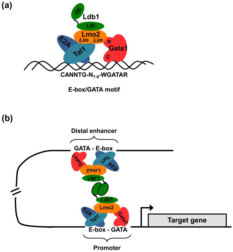
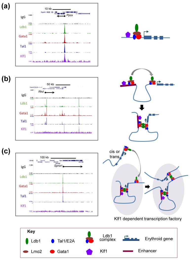
Elucidation of the genetic pathways that control red blood cell development has been a central goal of erythropoiesis research over the past decade. Notably, data from several recent studies have provided new insights into the regulation of erythroid gene transcription. Transcription profiling demonstrates that erythropoiesis is mainly controlled by a small group of lineage-restricted transcription factors [Gata binding protein 1 (Gata1), T cell acute lymphocytic leukemia 1 protein (Tal1), and Erythroid Kruppel-like factor (EKLF; henceforth referred to as Klf1)]. Binding-site mapping using ChIP-Seq indicates that most DNA-bound Gata1 and Tal1 proteins are contained within higher order complexes (Ldb1 complexes) that include the nuclear adapters Ldb1 and Lmo2. Ldb1 complexes regulate Klf1, and Ldb1 complex-binding sites frequently colocalize with Klf1 at erythroid genes and cis-regulatory elements, indicating strong functional synergy between Gata1, Tal1, and Klf1. Together with new data demonstrating that Ldb1 can mediate long-range promoter-enhancer interactions, these findings provide a foundation for the first comprehensive models of the global regulation of erythroid gene transcription.

Keywords: ChIP-Seq; Gata1; Klf1; Ldb1 complexes; Tal1; erythropoiesis; transcriptional regulation.


Figures

Figure 1
Figure 1. The structure and function of erythroid Ldb1-complexes
(a) Model of the core erythropoietic Ldb1-complex. The zinc finger DNA binding protein Gata1 and a heterodimer of the basic helix-loop-helix (b-HLH) proteins Tal1 and E2a bind to a paired E-box—WGATAR motif (W is A/T; R is A/G) with a restricted spacing of 7-9 bp. The dual LIM (Lin11, Isl-1 & Mec-3) domain protein Lmo2 bridges and associates with both Gata1 and the bHLH factors while the LIM interacting protein Ldb1 associates with Lmo2. Gata1 and Tal1 are described in more detail in Box 1. Ldb1 is a transcription co-factor widely expressed throughout embryonic and adult tissues. Ldb1 has no known enzymatic or nucleic acid-binding function, but rather it seems to act as an interface for specific protein interactions [29]. Ldb1 achieves this through its two predominant functional domains: an N-terminal self-association (dimerization) domain and a C-terminal region that interacts with the LIM domain that is common to a large family of proteins that have important roles in tissue development. Lmo2 is a small protein composed of two LIM domains and is expressed in a variety of tissues including hematopoietic precursors as well as many but not all hematopoietic lineages. In vitro binding studies have shown that association of Lmo2 with Tal1 increases the affinity of Tal1 to bind E2a and in turn, more stably bind to the E-box sequence [75]. Lmo2 also binds to the N-terminal zinc finger of Gata1, thereby forming a bridge between Tal1 and Gata1 [45]. Furthermore, it has been demonstrated that Gata1 can recruit additional proteins, such as Fog1 (Zfpm1), to the complex, through its N-terminal zinc finger [45]. A major function of the Ldb1-complex is to provide a stable scaffold through which the hematopoietic transcription factors Tal1 and Gata1 act together to regulate erythroid gene transcription. (b) An illustration of Ldb1-mediated juxtaposition of two Ldb1-complexes bound to DNA at sites far apart from each other. Ldb1 can dimerize through its self-association domain facilitating DNA looping and juxtaposition of two Ldb1-complexes. This property of Ldb1 suggests a model whereby enhancers can communicate with distal promoters (in cis or possibly in trans) via Ldb1-complex mediated association. The Ldb1 self-association domain can also form trimeric structures as well as dimers and these types of higher order structures are likely relevant in instances where multiple Ldb1-complexes are assembled near a gene (e.g. the Hbb locus) [30].
Figure 2
Figure 2. Hypothetical models for the cooperation of Ldb1-complexes and Klf1 in the regulation of erythroid gene transcription
Genome-wide mapping of Ldb1-complex and Klf1 binding by ChIP-Seq suggests several possible mechanisms by which Klf1 functions in concert with the Ldb1-complex to activate erythroid genes known to be dependent on both Klf1 and Ldb1 for their expression. Images on the left represent raw ChIP-Seq read data for Ldb1, Gata1, Tal1, and Klf1 [8, 25] transformed into a density plot for each factor and loaded into the UCSC genome browser as a custom track. Models corresponding to the binding profiles are depicted on the right. (a) Left, example of an erythroid gene (Ppox) where an Ldb1-complex and Klf1 bind in close proximity to each other and to the transcription start site of a gene to directly activate transcription. Right, model depicting the direct regulation of Ppox transcription by the Ldb1-complex and Klf1. (b) Left, example of an erythroid gene (Rhd) where an Ldb1-complex but not Klf1 binds at the promoter and where both an Ldb1-complex and Klf1 bind to a distal enhancer. Right, model depicting the recruitment of Klf1 to the Rhd promoter through dimerization of the Ldb1 self-association domain. (c) Left, example of an erythroid gene (Spna1) where Ldb1-complex binding is detected at the gene but Klf1 binding is not detected within 100 kilobases of the gene. Right, model depicts Ldb1-complex mediated recruitment of Spna1 to a transcriptional hub near the Hbb gene. In this model, the transcriptional hub serves as a nexus where Klf1-dependent genes are brought into locations of direct Klf1 recruitment through the self-interactions of Ldb1-complexes.

References

    1. Pevny L, et al. Erythroid differentiation in chimaeric mice blocked by a targeted mutation in the gene for transcription factor GATA-1. Nature. 1991;349:257–260. - PubMed
    1. Schlaeger TM, et al. Tie2Cre-mediated gene ablation defines the stem-cell leukemia gene (SCL/tal1)-dependent window during hematopoietic stem-cell development. Blood. 2005;105:3871–3874. - PubMed
    1. Robb L, et al. Absence of yolk sac hematopoiesis from mice with a targeted disruption of the scl gene. Proceedings of the National Academy of Sciences of the United States of America. 1995;92:7075–7079. - PMC - PubMed
    1. Porcher C, et al. The T cell leukemia oncoprotein SCL/tal-1 is essential for development of all hematopoietic lineages. Cell. 1996;86:47–57. - PubMed
    1. Nuez B, et al. Defective haematopoiesis in fetal liver resulting from inactivation of the EKLF gene. Nature. 1995;375:316–318. - PubMed

Publication types

MeSH terms

Substances