

Pituitary gland development and disease: from stem cell to hormone production
- PMID: 24290346
- PMCID: PMC4039019
- DOI: 10.1016/B978-0-12-416021-7.00001-8
Pituitary gland development and disease: from stem cell to hormone production
Abstract
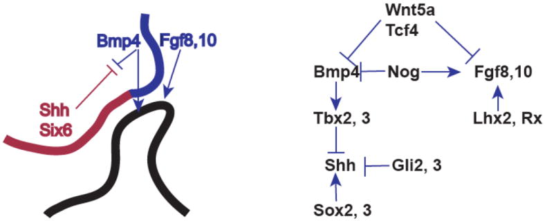
Many aspects of pituitary development have become better understood in the past two decades. The signaling pathways regulating pituitary growth and shape have emerged, and the balancing interactions between the pathways are now appreciated. Markers for multipotent progenitor cells are being identified, and signature transcription factors have been discovered for most hormone-producing cell types. We now realize that pulsatile hormone secretion involves a 3D integration of cellular networks. About a dozen genes are known to cause pituitary hypoplasia when mutated due to their essential roles in pituitary development. Similarly, a few genes are known that predispose to familial endocrine neoplasia, and several genes mutated in sporadic pituitary adenomas are documented. In the next decade, we anticipate gleaning a deeper appreciation of these processes at the molecular level, insight into the development of the hypophyseal portal blood system, and evolution of better therapeutics for congenital and acquired hormone deficiencies and for common craniopharyngiomas and pituitary adenomas.
Keywords: Adenohypophysis; Anterior pituitary; Neural ectoderm; Organizing center; Rathke’s pouch; Stem cell.
© 2013 Elsevier Inc. All rights reserved.
Figures
References
-
- Schwind JL. The development of the hypophysis cerebri of the albino rat. Amer J Anat. 1928;41:295–315.
-
- Rathke MH. Entwicklungsgeschichte der Natter (Coluber natrix) Königsberg Bornträger; 1839.
-
- Couly GF, Le Douarin NM. Mapping of the early neural primordium in quail-chick chimeras. I. Developmental relationships between placodes, facial ectoderm, and prosencephalon. Dev Biol. 1985;110(2):422–39. - PubMed
-
- Gleiberman AS, Fedtsova NG, Rosenfeld MG. Tissue interactions in the induction of anterior pituitary: role of the ventral diencephalon, mesenchyme, and notochord. Dev Biol. 1999;213(2):340–53. - PubMed
-
- Takuma N, Sheng HZ, Furuta Y, et al. Formation of Rathke’s pouch requires dual induction from the diencephalon. Development. 1998;125(23):4835–40. - PubMed
Publication types
MeSH terms
Substances
Grants and funding
LinkOut - more resources
Full Text Sources
Other Literature Sources
Medical
Miscellaneous
