. 2013 Dec 12;5(5):1178-86.
doi: 10.1016/j.celrep.2013.10.049. Epub 2013 Nov 27.

Hexanucleotide repeats in ALS/FTD form length-dependent RNA foci, sequester RNA binding proteins, and are neurotoxic

Affiliations

Hexanucleotide repeats in ALS/FTD form length-dependent RNA foci, sequester RNA binding proteins, and are neurotoxic

Youn-Bok Lee et al. Cell Rep. .

Abstract

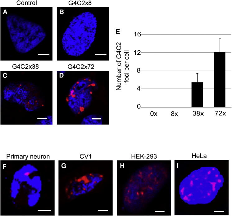
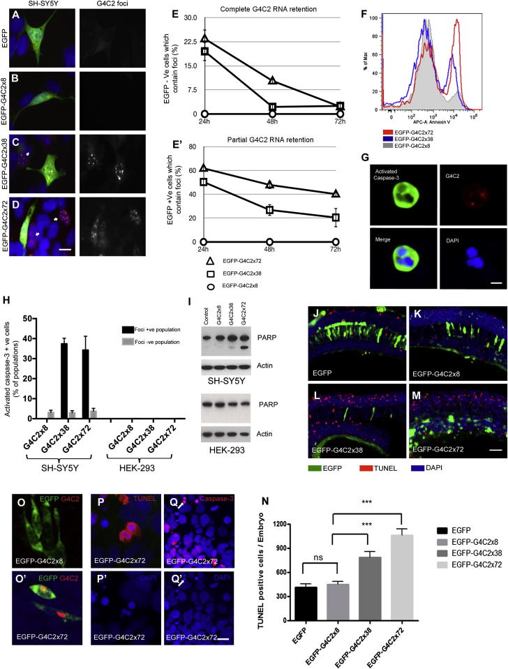
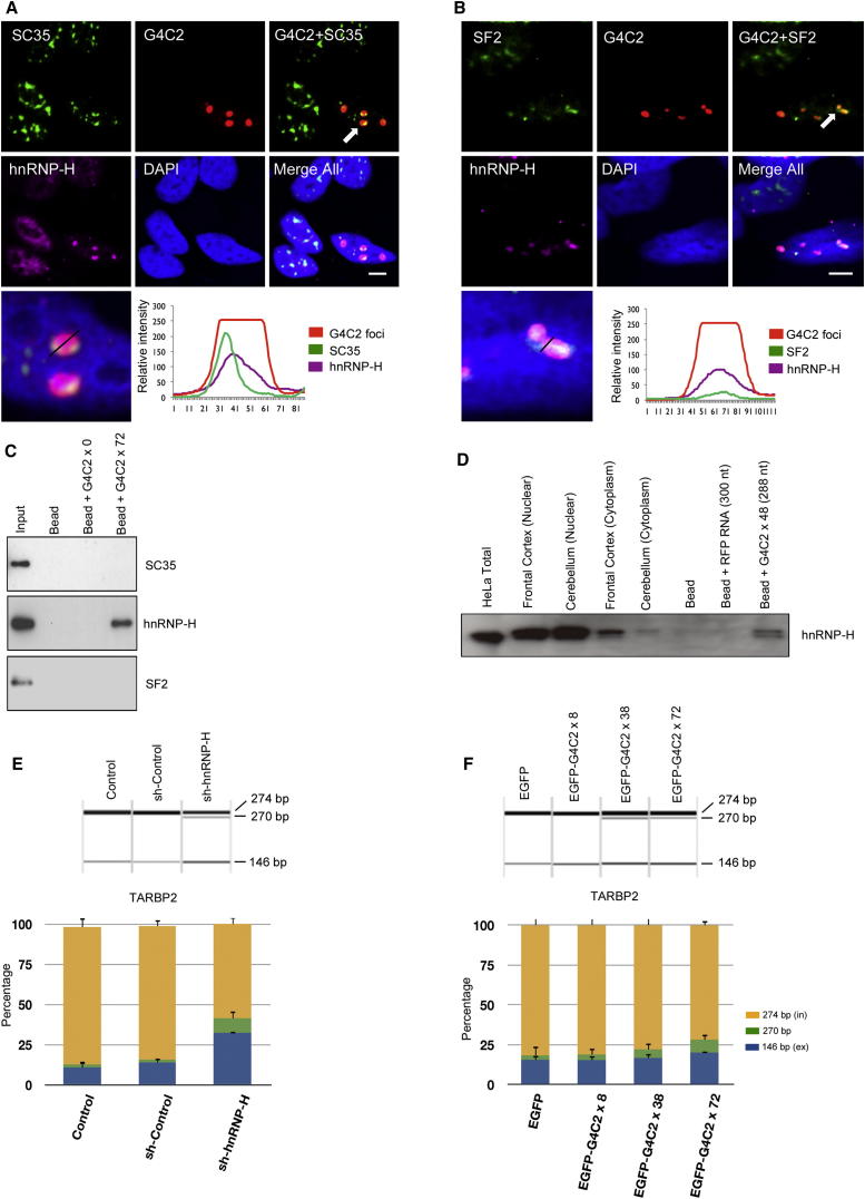
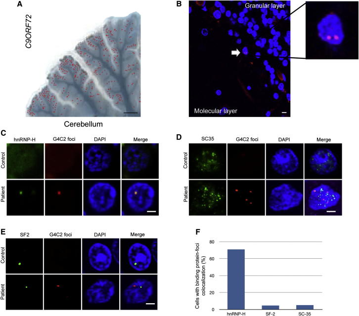
The GGGGCC (G4C2) intronic repeat expansion within C9ORF72 is the most common genetic cause of amyotrophic lateral sclerosis (ALS) and frontotemporal dementia (FTD). Intranuclear neuronal RNA foci have been observed in ALS and FTD tissues, suggesting that G4C2 RNA may be toxic. Here, we demonstrate that the expression of 38× and 72× G4C2 repeats form intranuclear RNA foci that initiate apoptotic cell death in neuronal cell lines and zebrafish embryos. The foci colocalize with a subset of RNA binding proteins, including SF2, SC35, and hnRNP-H in transfected cells. Only hnRNP-H binds directly to G4C2 repeats following RNA immunoprecipitation, and only hnRNP-H colocalizes with 70% of G4C2 RNA foci detected in C9ORF72 mutant ALS and FTD brain tissues. We show that expanded G4C2 repeats are potently neurotoxic and bind hnRNP-H and other RNA binding proteins. We propose that RNA toxicity and protein sequestration may disrupt RNA processing and contribute to neurodegeneration.


Figures

None
Graphical abstract
Figure 1
Figure 1
Expanded G4C2 Repeats Form Intranuclear RNA Foci (A–D) SH-SY5Y cells were transfected with various length G4C2 (0, 8, 38, 72 repeat) plasmids and analyzed 24 hr after transfection by RNA FISH using a Cy3-labeled (G2C4)×8 RNA probe. (E) The mean number of G4C2 foci was counted in 50 cells. (F–I) Primary hippocampal mouse neurons (F), CV1 (G), HEK293 (H), and HeLa (I) cells were transfected with a plasmid expressing G4C2 72× repeats; foci were detected by FISH (red) and nuclei were stained with DAPI (blue). All cell types transfected with 38× and 72× repeats showed foci (scale bar represents 3 μm). See also Figure S1.
Figure 2
Figure 2
Loss of G4C2 Foci-Positive Cells Is Due to Apoptotic Cell Death in Culture and In Vivo (A–D) SH-SY5Y cells were transfected with EGFP-tagged G4C2 constructs. G4C2 RNA foci (red) were found only in cells transfected with EGFP-G4C2 38× and EGFP-G4C2 72× repeats. Many cells with RNA foci were negative for EGFP (arrows in C and D), implying near-complete nuclear retention. Nuclei were stained with DAPI (blue) (scale bar represents 10 μm). (E) The percentage of cells with G4C2 foci by FISH that did not express EGFP were counted at 24, 48, and 72 hr posttransfection (E), as were foci-positive cells that did express EGFP (E’). Foci-positive cells declined in number, most markedly in those showing greater nuclear retention (EGFP−). In all experiments, a total of 250 cells from three independent transfections were counted, and results are presented as mean ± SD. (F) EGFP-tagged G4C2 plasmids were transfected into SH-SY5Y cells, stained with the early apoptosis marker Annexin V, and analyzed by FACS. Cells expressing 38× and 72× repeats showed 3- or 5-fold higher levels of Annexin V, respectively, than did cells expressing 8× repeats. (G) G4C2 foci-positive SH-SY5Y cells were found positive for activated caspase-3 (scale bar represents 5 μm). (H) G4C2 RNA foci-positive SH-SY5Y cells, but not HEK293 cells, express activated caspase-3. Caspase-3 activation was scored in 250 foci-positive cells per coverslip, and three coverslips were analyzed per experiment. The background level of active caspase-3 was estimated by counting the frequency of active caspase-3-positive cells in the foci-negative population. (I) Western blot of PARP cleavage in control or G4C2-transfected SH-SY5Y and HEK293 cells. Actin was used as a loading control. (J–M) Apoptotic cell death was analyzed by TUNEL staining in zebrafish prim-5 embryos injected with plasmids mosaically expressing EGFP (J), EGFP-G4C2 8× (K), EGFP-G4C2 38× (L), or EGFP-G4C2 72× (M). The number of TUNEL-positive cells (red) increased in the embryos injected with 38× and 72× repeats (L and M) (scale bar represents 200 μm). (N) Quantification of all embryos (n = 5) from three independent experiments is presented. Error bars show the standard error for each sample, and p values (p < 0.0001, ∗∗∗) are also determined. (O) G4C2 RNA foci (red) were found only in zebrafish embryo cells injected with 72×, but not with 8× repeats. EGFP expression is green. (P and Q) High-resolution images of TUNEL-positive (P and P′) and active caspse-3 (Q and Q′) from zebrafish embryo cells injected with 72× (scale bar represents 10 μm). Nuclear staining is blue. See also Figure S2.
Figure 3
Figure 3
SC35, SF2, and hnRNP-H Colocalize with G4C2 Nuclear Foci, but hnRNP-H Binds to G4C2 RNA Transcripts (A–B) SH-SY5Y cells were transfected with a plasmid expressing 72× repeats and probed 24 hr after transfection for G4C2 by FISH and immunocytochemistry (ICC). (A and B) Endogenous SC35 (A) and SF2 (B) were detected by ICC using a Dylight-488-labeled secondary antibody. hnRNP-H was simultaneously detected by ICC, using a Dylight-649-labeled secondary antibody. The intensity of endogenous SC35, SF2, hnRNP-H, and G4C2 foci were analyzed by Leica line profile tools (scale bar represents 5 μm). (C) Biotin-labeled G4C2×72 RNA transcripts were synthesized and used for RNA pull-down of SH-SH5Y lysates; G4C2×0 transcripts were used as control. Partial colocalization of SC35 and SF2 is seen with the RNA foci, whereas hnRNP-H shows very close colocalization. Only hnRNP-H coprecipitated with G4C2×72 RNA. (D) Biotin aptamer-labeled G4C2×48 RNA transcripts were synthesized and used for RNA pull-down of rat brain lysates (Supplemental Experimental Procedures); RNA of equivalent length to G4C2×48 (300 nt) was used as a control. hnRNP-H coprecipitated with G4C2×48 RNA. (E) Splicing assay of TARBP2 exon 7 shows that G4C2 RNA sequestration of hnRNP-H impairs RNA processing. Comparison of hnRNP H knockdown and control SH-SY5Y cells validates that hnRNP H promotes inclusion of TARBP2 exon 7. (F) Analysis of TARBP2 exon 7 splicing in SH-SY5Y cells stably transfected with C9ORF72 repeats. Inclusion of TARBP2 exon 7 is decreased in cells expressing G4C2 ×72 but is not affected in cells expressing lower repeat numbers. PCR products including (in) or excluding (ex) the regulated alternative exon are marked on the right. Average quantification values of exon inclusion (yellow) and exclusion (blue) are shown. Error bars show SD of three replicates. To test significance, the ratio between exon 7 inclusion and exclusion was calculated and tested by one-way ANOVA and Tukey’s honestly significant difference test. See also Figure S3.
Figure 4
Figure 4
Intranuclear Neuronal RNA Foci in C9ORF72 Mutant ALS and FTD Brain Tissues Colocalize Very Closely with hnRNP-H (A) Image of mutant C9ORF72 patient cerebellum overlaid with the location of neurons containing G4C2 RNA foci (red dots) (scale bar represents 4 mm). (B) G4C2 RNA foci-positive neurons (white arrow) were observed between the granular and molecular layer of the cerebellum (scale bar represents 10 μm). (C–E) FISH and ICC were performed for hnRNP-H (C), SC35 (D), and SF2 (E), with a G4C2 mutation-negative ALS case used as control (scale bar represents 3 μm). (F) The percentage of foci that colocalized with hnRNP-H, SF2, and SC-35 were counted (n = 50 cells). Of the three RNA binding proteins that colocalized with foci in transfected cells, only hnRNP-H shows a striking degree of overlap for 70% of all foci in the cerebellum. See also Figure S4.

Similar articles

Cited by

References

    1. Al-Sarraj S., King A., Troakes C., Smith B., Maekawa S., Bodi I., Rogelj B., Al-Chalabi A., Hortobágyi T., Shaw C.E. p62 positive, TDP-43 negative, neuronal cytoplasmic and intranuclear inclusions in the cerebellum and hippocampus define the pathology of C9orf72-linked FTLD and MND/ALS. Acta Neuropathol. 2011;122:691–702. - PubMed
    1. Chou M.Y., Rooke N., Turck C.W., Black D.L. hnRNP H is a component of a splicing enhancer complex that activates a c-src alternative exon in neuronal cells. Mol. Cell. Biol. 1999;19:69–77. - PMC - PubMed
    1. DeJesus-Hernandez M., Mackenzie I.R., Boeve B.F., Boxer A.L., Baker M., Rutherford N.J., Nicholson A.M., Finch N.A., Flynn H., Adamson J. Expanded GGGGCC hexanucleotide repeat in noncoding region of C9ORF72 causes chromosome 9p-linked FTD and ALS. Neuron. 2011;72:245–256. - PMC - PubMed
    1. Fratta P., Mizielinska S., Nicoll A.J., Zloh M., Fisher E.M., Parkinson G., Isaacs A.M. C9orf72 hexanucleotide repeat associated with amyotrophic lateral sclerosis and frontotemporal dementia forms RNA G-quadruplexes. Sci Rep. 2012;2:1016. - PMC - PubMed
    1. Hirth F. Drosophila melanogaster in the study of human neurodegeneration. CNS Neurol. Disord. Drug Targets. 2010;9:504–523. - PMC - PubMed

Publication types

MeSH terms